4WTH | pdb_00004wth

Ataxin-3 Carboxy Terminal Region - Crystal C2 (triclinic)

  • Classification: TRANSCRIPTION
  • Organism(s): Escherichia coli, Homo sapiens
  • Expression System: Escherichia coli BL21(DE3)
  • Mutation(s): No 

  • Deposited: 2014-10-30 Released: 2016-03-09 
  • Deposition Author(s): Zhemkov, V.A., Kim, M.
  • Funding Organization(s): National Institutes of Health/National Institute of Neurological Disorders and Stroke (NIH/NINDS), National Ataxia Foundation, Russian Ministry of Science and Education, Russian Scientific Fund

Experimental Data Snapshot

  • Method: X-RAY DIFFRACTION
  • Resolution: 2.25 Å
  • R-Value Free: 
    0.250 (Depositor), 0.214 (DCC) 
  • R-Value Work: 
    0.202 (Depositor) 
  • R-Value Observed: 
    0.204 (Depositor) 

Starting Model: experimental
View more details

wwPDB Validation 3D Report Full Report

Validation slider image for 4WTH

This is version 2.1 of the entry. See complete history

Literature

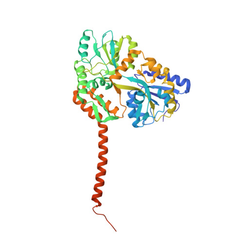
The 2.2-Angstrom resolution crystal structure of the carboxy-terminal region of ataxin-3.

Zhemkov, V.A.Kulminskaya, A.A.Bezprozvanny, I.B.Kim, M.

(2016) FEBS Open Bio 6: 168-178

  • DOI: https://doi.org/10.1002/2211-5463.12029
  • Primary Citation Related Structures: 
    4WTH, 4YS9

  • PubMed Abstract: 

    An expansion of polyglutamine (polyQ) sequence in ataxin-3 protein causes spinocerebellar ataxia type 3, an inherited neurodegenerative disorder. The crystal structure of the polyQ-containing carboxy-terminal fragment of human ataxin-3 was solved at 2.2-Å resolution. The Atxn3 carboxy-terminal fragment including 14 glutamine residues adopts both random coil and α-helical conformations in the crystal structure. The polyQ sequence in α-helical structure is stabilized by intrahelical hydrogen bonds mediated by glutamine side chains. The intrahelical hydrogen-bond interactions between glutamine side chains along the axis of the polyQ α-helix stabilize the secondary structure. Analysis of this structure furthers our understanding of the polyQ-structural characteristics that likely underlie the pathogenesis of polyQ-expansion disorders.


  • Organizational Affiliation
    • Laboratory of Molecular Neurodegeneration St Petersburg State Polytechnical University Russia; Laboratory of Enzymology National Research Center «Kurchatov Institute»B.P. Konstantinov Petersburg Nuclear Physics Institute Gatchina Russia.

Macromolecule Content 

  • Total Structure Weight: 98.23 kDa 
  • Atom Count: 6,458 
  • Modeled Residue Count: 809 
  • Deposited Residue Count: 882 
  • Unique protein chains: 1

Macromolecules

Find similar proteins by:|  3D Structure
Entity ID: 1
MoleculeChains  Sequence LengthOrganismDetailsImage
Maltose-binding periplasmic protein, Ataxin-3 chimera
A, B
441Escherichia coliHomo sapiensMutation(s): 0 
Gene Names: malEZ5632ECs5017ATXN3ATX3MJDMJD1SCA3
EC: 3.4.19.12
UniProt & NIH Common Fund Data Resources
Find proteins for P0AEX9 (Escherichia coli (strain K12))
Explore P0AEX9 
Go to UniProtKB:  P0AEX9
Find proteins for P54252 (Homo sapiens)
Explore P54252 
Go to UniProtKB:  P54252
PHAROS:  P54252
GTEx:  ENSG00000066427 
Entity Groups
Sequence Clusters30% Identity50% Identity70% Identity90% Identity95% Identity100% Identity
UniProt GroupsP0AEX9P54252
Sequence Annotations
Expand
Reference Sequence

Oligosaccharides

Help  
Entity ID: 2
MoleculeChains Length2D Diagram GlycosylationD Interactions
alpha-D-glucopyranose-(1-4)-alpha-D-glucopyranose
C, D
2N/A
Glycosylation Resources
GlyTouCan: G07411ON
GlyCosmos: G07411ON

Biologically Interesting Molecules (External Reference) 

1 Unique

Experimental Data & Validation

Experimental Data

  • Method: X-RAY DIFFRACTION
  • Resolution: 2.25 Å
  • R-Value Free:  0.250 (Depositor), 0.214 (DCC) 
  • R-Value Work:  0.202 (Depositor) 
  • R-Value Observed: 0.204 (Depositor) 
Space Group: P 1
Unit Cell:
Length ( Å )Angle ( ˚ )
a = 49.096α = 90
b = 59.786β = 89.99
c = 77.79γ = 87.5
Software Package:
Software NamePurpose
REFMACrefinement
HKL-2000data collection
HKL-2000data reduction
SCALEPACKdata scaling
PHASERphasing
Cootmodel building

Structure Validation

View Full Validation Report


Entry History 

& Funding Information

Deposition Data


Funding OrganizationLocationGrant Number
National Institutes of Health/National Institute of Neurological Disorders and Stroke (NIH/NINDS)United StatesR01NS074376
National Institutes of Health/National Institute of Neurological Disorders and Stroke (NIH/NINDS)United StatesR01NS056224
National Ataxia FoundationUnited StatesTranslational Award
Russian Ministry of Science and EducationRussian Federation17.1360.2014/K
Russian Scientific FundRussian Federation14-25-00024
Russian Ministry of Science and EducationRussian Federation11.G34.31.0056

Revision History  (Full details and data files)

  • Version 1.0: 2016-03-09
    Type: Initial release
  • Version 1.1: 2016-03-16
    Changes: Database references
  • Version 1.2: 2017-09-06
    Changes: Author supporting evidence, Derived calculations
  • Version 1.3: 2018-04-18
    Changes: Data collection, Database references
  • Version 1.4: 2019-12-18
    Changes: Author supporting evidence
  • Version 2.0: 2020-07-29
    Type: Remediation
    Reason: Carbohydrate remediation
    Changes: Atomic model, Data collection, Derived calculations, Non-polymer description, Structure summary
  • Version 2.1: 2023-09-27
    Changes: Data collection, Database references, Refinement description, Structure summary