4WSI | pdb_00004wsi

Crystal Structure of PALS1/Crb complex


Experimental Data Snapshot

  • Method: X-RAY DIFFRACTION
  • Resolution: 2.95 Å
  • R-Value Free: 
    0.235 (Depositor), 0.262 (DCC) 
  • R-Value Work: 
    0.207 (Depositor) 
  • R-Value Observed: 
    0.208 (Depositor) 

wwPDB Validation 3D Report Full Report

Validation slider image for 4WSI

This is version 1.2 of the entry. See complete history

Literature

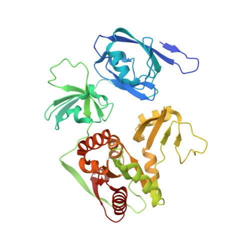
Structure of Crumbs tail in complex with the PALS1 PDZ-SH3-GK tandem reveals a highly specific assembly mechanism for the apical Crumbs complex.

Li, Y.Wei, Z.Yan, Y.Wan, Q.Du, Q.Zhang, M.

(2014) Proc Natl Acad Sci U S A 111: 17444-17449

  • DOI: https://doi.org/10.1073/pnas.1416515111
  • Primary Citation Related Structures: 
    4WSI

  • PubMed Abstract: 

    The Crumbs (Crb) complex, formed by Crb, PALS1, and PATJ, is evolutionarily conserved in metazoans and acts as a master cell-growth and -polarity regulator at the apical membranes in polarized epithelia. Crb intracellular functions, including its direct binding to PALS1, are mediated by Crb's highly conserved 37-residue cytoplasmic tail. However, the mechanistic basis governing the highly specific Crb-PALS1 complex formation is unclear, as reported interaction between the Crb tail (Crb-CT) and PALS1 PSD-95/DLG/ZO-1 (PDZ) domain is weak and promiscuous. Here we have discovered that the PDZ-Src homolgy 3 (SH3)-Guanylate kinase (GK) tandem of PALS1 binds to Crb-CT with a dissociation constant of 70 nM, which is ∼ 100-fold stronger than the PALS1 PDZ-Crb-CT interaction. The crystal structure of the PALS1 PDZ-SH3-GK-Crb-CT complex reveals that PDZ-SH3-GK forms a structural supramodule with all three domains contributing to the tight binding to Crb. Mutations disrupting the tertiary interactions of the PDZ-SH3-GK supramodule weaken the PALS1-Crb interaction and compromise PALS1-mediated polarity establishment in Madin-Darby canine kidney (MDCK) cysts. We further show that specific target binding of other members of membrane-associated guanylate kinases (MAGUKs) (e.g., CASK binding to neurexin) also requires the presence of their PDZ-SH3-GK tandems.


  • Organizational Affiliation
    • Division of Life Science, State Key Laboratory of Molecular Neuroscience and.

Macromolecule Content 

  • Total Structure Weight: 99.44 kDa 
  • Atom Count: 6,042 
  • Modeled Residue Count: 767 
  • Deposited Residue Count: 874 
  • Unique protein chains: 2

Macromolecules

Find similar proteins by:|  3D Structure
Entity ID: 1
MoleculeChains  Sequence LengthOrganismDetailsImage
MAGUK p55 subfamily member 5
A, B
394Homo sapiensMutation(s): 0 
Gene Names: MPP5
UniProt & NIH Common Fund Data Resources
Find proteins for Q8N3R9 (Homo sapiens)
Explore Q8N3R9 
Go to UniProtKB:  Q8N3R9
PHAROS:  Q8N3R9
GTEx:  ENSG00000072415 
Entity Groups
Sequence Clusters30% Identity50% Identity70% Identity90% Identity95% Identity100% Identity
UniProt GroupQ8N3R9
Sequence Annotations
Expand
Reference Sequence
Find similar proteins by:|  3D Structure
Entity ID: 2
MoleculeChains  Sequence LengthOrganismDetailsImage
Protein crumbsC [auth X],
D [auth Y]
43Drosophila melanogasterMutation(s): 0 
Gene Names: crbCG6383
UniProt
Find proteins for P10040 (Drosophila melanogaster)
Explore P10040 
Go to UniProtKB:  P10040
Entity Groups
Sequence Clusters30% Identity50% Identity70% Identity90% Identity95% Identity100% Identity
UniProt GroupP10040
Sequence Annotations
Expand
Reference Sequence

Experimental Data & Validation

Experimental Data

  • Method: X-RAY DIFFRACTION
  • Resolution: 2.95 Å
  • R-Value Free:  0.235 (Depositor), 0.262 (DCC) 
  • R-Value Work:  0.207 (Depositor) 
  • R-Value Observed: 0.208 (Depositor) 
Space Group: P 41 21 2
Unit Cell:
Length ( Å )Angle ( ˚ )
a = 111.928α = 90
b = 111.928β = 90
c = 223.065γ = 90
Software Package:
Software NamePurpose
Blu-Icedata collection
HKL-2000data processing
PHENIXphasing
Cootmodel building
PHENIXrefinement
PDB_EXTRACTdata extraction
HKL-2000data scaling

Structure Validation

View Full Validation Report


Entry History 

Deposition Data

Revision History  (Full details and data files)

  • Version 1.0: 2014-11-26
    Type: Initial release
  • Version 1.1: 2015-02-11
    Changes: Database references
  • Version 1.2: 2024-03-20
    Changes: Data collection, Database references, Derived calculations, Source and taxonomy