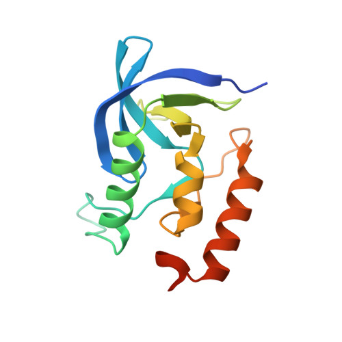
4WOR

Staphylococcal nuclease in complex with Ca2+ and thymidine-3'-5'-diphosphate (pdTp) at room temperature


Experimental Data Snapshot

  • Method: X-RAY DIFFRACTION
  • Resolution: 1.60 Å
  • R-Value Free: 0.204 
  • R-Value Work: 0.180 
  • R-Value Observed: 0.181 

wwPDB Validation   3D Report Full Report


Ligand Structure Quality Assessment 


This is version 2.2 of the entry. See complete history


Literature

Three-dimensional diffuse x-ray scattering from crystals of Staphylococcal nuclease.

Wall, M.E.Ealick, S.E.Gruner, S.M.

(1997) Proc Natl Acad Sci U S A : 6180-6184

  • DOI: https://doi.org/10.1073/pnas.94.12.6180
  • Primary Citation of Related Structures:  
    4WOR

  • PubMed Abstract: 

    We have developed methods for obtaining and characterizing three-dimensional maps of the reciprocal-space distribution of diffuse x-ray scattering from protein crystals, and have used the methods to study the nature of disorder in crystals of Staphylococcal nuclease. Experimentally obtained maps are 99.5% complete in the reciprocal-space resolution range of 10 A-2.5 A, show symmetry consistent with the P41 space group of the unit cell, and are highly reproducible. Quantitative comparisons of the data with three-dimensional simulations imply liquid-like motions of the protein [Caspar, D. L. D., Clarage, J., Salunke, D. M. & Clarage, M. (1988) Nature (London) 332, 659-662], with a correlation length of 10 A and a root-mean-square displacement of 0.36 A.


  • Organizational Affiliation

    Department of Physics, Princeton University, Princeton, NJ 08544, USA.


Macromolecules
Find similar proteins by:  (by identity cutoff)  |  3D Structure
Entity ID: 1
MoleculeChains Sequence LengthOrganismDetailsImage
Thermonuclease149Staphylococcus aureusMutation(s): 0 
Gene Names: nuc
EC: 3.1.31.1
UniProt
Find proteins for P00644 (Staphylococcus aureus)
Explore P00644 
Go to UniProtKB:  P00644
Entity Groups  
Sequence Clusters30% Identity50% Identity70% Identity90% Identity95% Identity100% Identity
UniProt GroupP00644
Sequence Annotations
Expand
  • Reference Sequence
Small Molecules
Ligands 2 Unique
IDChains Name / Formula / InChI Key2D Diagram3D Interactions
THP
Query on THP

Download Ideal Coordinates CCD File 
C [auth A]THYMIDINE-3',5'-DIPHOSPHATE
C10 H16 N2 O11 P2
CSNCBOPUCJOHLS-XLPZGREQSA-N
CA
Query on CA

Download Ideal Coordinates CCD File 
B [auth A]CALCIUM ION
Ca
BHPQYMZQTOCNFJ-UHFFFAOYSA-N
Experimental Data & Validation

Experimental Data

  • Method: X-RAY DIFFRACTION
  • Resolution: 1.60 Å
  • R-Value Free: 0.204 
  • R-Value Work: 0.180 
  • R-Value Observed: 0.181 
  • Space Group: P 41
Unit Cell:
Length ( Å )Angle ( ˚ )
a = 48.499α = 90
b = 48.499β = 90
c = 63.43γ = 90
Software Package:
Software NamePurpose
PHENIXrefinement

Structure Validation

View Full Validation Report


Ligand Structure Quality Assessment 


Entry History & Funding Information

Deposition Data


Funding OrganizationLocationGrant Number
Department of Energy (DOE, United States)United States--

Revision History  (Full details and data files)

  • Version 1.0: 2014-10-29
    Type: Initial release
  • Version 2.0: 2017-09-20
    Changes: Atomic model, Author supporting evidence, Derived calculations, Other, Source and taxonomy
  • Version 2.1: 2019-12-04
    Changes: Author supporting evidence
  • Version 2.2: 2023-09-27
    Changes: Data collection, Database references, Derived calculations, Refinement description