4WO5 | pdb_00004wo5

Crystal structure of a BRAF kinase domain monomer


Experimental Data Snapshot

  • Method: X-RAY DIFFRACTION
  • Resolution: 2.83 Å
  • R-Value Free: 
    0.260 (Depositor), 0.268 (DCC) 
  • R-Value Work: 
    0.193 (Depositor), 0.201 (DCC) 
  • R-Value Observed: 
    0.196 (Depositor) 

Starting Model: experimental
View more details

wwPDB Validation 3D Report Full Report

Validation slider image for 4WO5

Ligand Structure Quality Assessment 


This is version 1.4 of the entry. See complete history

Literature

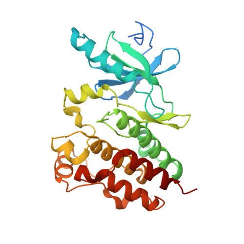
Crystal structure of a BRAF kinase domain monomer explains basis for allosteric regulation.

Thevakumaran, N.Lavoie, H.Critton, D.A.Tebben, A.Marinier, A.Sicheri, F.Therrien, M.

(2015) Nat Struct Mol Biol 22: 37-43

  • DOI: https://doi.org/10.1038/nsmb.2924
  • Primary Citation Related Structures: 
    4WO5

  • PubMed Abstract: 

    Reported RAF kinase domain structures adopt a side-to-side dimer configuration reflective of an 'on' state that underpins an allosteric mechanism of regulation. Atomic details of the monomer 'off' state have been elusive. Reinspection of the BRAF kinase domain structures revealed that sulfonamide inhibitors induce features of an off state, primarily a laterally displaced helix αC stabilized by the activation segment helix 1 (AS-H1). These features correlated with the ability of sulfonamides to disrupt human BRAF homodimers in cells, in vitro and in crystals yielding a structure of BRAF in a monomer state. The crystal structure revealed exaggerated, nonproductive positions of helix αC and AS-H1, the latter of which is the target of potent BRAF oncogenic mutations. Together, this work provides formal proof of an allosteric link between the RAF dimer interface, the activation segment and the catalytic infrastructure.


  • Organizational Affiliation
    • 1] Lunenfeld-Tanenbaum Research Institute, Mount Sinai Hospital, Toronto, Ontario, Canada. [2] Department of Biochemistry, University of Toronto, Toronto, Ontario, Canada.

Macromolecule Content 

  • Total Structure Weight: 69.06 kDa 
  • Atom Count: 4,109 
  • Modeled Residue Count: 523 
  • Deposited Residue Count: 600 
  • Unique protein chains: 1

Macromolecules

Find similar proteins by:|  3D Structure
Entity ID: 1
MoleculeChains  Sequence LengthOrganismDetailsImage
Serine/threonine-protein kinase B-raf
A, B
300Homo sapiensMutation(s): 16 
Gene Names: BRAFBRAF1RAFB1
EC: 2.7.11.1
UniProt & NIH Common Fund Data Resources
Find proteins for P15056 (Homo sapiens)
Explore P15056 
Go to UniProtKB:  P15056
PHAROS:  P15056
GTEx:  ENSG00000157764 
Entity Groups
Sequence Clusters30% Identity50% Identity70% Identity90% Identity95% Identity100% Identity
UniProt GroupP15056
Sequence Annotations
Expand
Reference Sequence

Small Molecules

Ligands 1 Unique
IDChains Name / Formula / InChI Key2D Diagram3D Interactions
324

Query on 324


Download:Ideal Coordinates CCD File
C [auth A],
D [auth B]
N-{3-[(5-chloro-1H-pyrrolo[2,3-b]pyridin-3-yl)carbonyl]-2,4-difluorophenyl}propane-1-sulfonamide
C17 H14 Cl F2 N3 O3 S
YZDJQTHVDDOVHR-UHFFFAOYSA-N

Experimental Data & Validation

Experimental Data

  • Method: X-RAY DIFFRACTION
  • Resolution: 2.83 Å
  • R-Value Free:  0.260 (Depositor), 0.268 (DCC) 
  • R-Value Work:  0.193 (Depositor), 0.201 (DCC) 
  • R-Value Observed: 0.196 (Depositor) 
Space Group: I 2 2 2
Unit Cell:
Length ( Å )Angle ( ˚ )
a = 65.61α = 90
b = 72.748β = 90
c = 243.338γ = 90
Software Package:
Software NamePurpose
BUSTERrefinement

Structure Validation

View Full Validation Report


Ligand Structure Quality Assessment 


Entry History 

Deposition Data

Revision History  (Full details and data files)

  • Version 1.0: 2014-12-03
    Type: Initial release
  • Version 1.1: 2014-12-17
    Changes: Database references
  • Version 1.2: 2015-01-14
    Changes: Database references
  • Version 1.3: 2015-02-04
    Changes: Derived calculations
  • Version 1.4: 2023-09-27
    Changes: Data collection, Database references, Derived calculations, Other, Refinement description, Source and taxonomy