4WNA

Structure of the Nitrogenase MoFe Protein from Azotobacter vinelandii Pressurized with Xenon


Experimental Data Snapshot

  • Method: X-RAY DIFFRACTION
  • Resolution: 2.00 Å
  • R-Value Free: 0.219 
  • R-Value Work: 0.167 
  • R-Value Observed: 0.170 

wwPDB Validation   3D Report Full Report


Ligand Structure Quality Assessment 


This is version 1.5 of the entry. See complete history


Literature

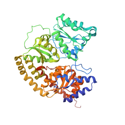
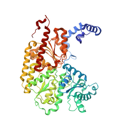
Substrate Pathways in the Nitrogenase MoFe Protein by Experimental Identification of Small Molecule Binding Sites.

Morrison, C.N.Hoy, J.A.Zhang, L.Einsle, O.Rees, D.C.

(2015) Biochemistry 54: 2052-2060

  • DOI: https://doi.org/10.1021/bi501313k
  • Primary Citation of Related Structures:  
    4WN9, 4WNA

  • PubMed Abstract: 

    In the nitrogenase molybdenum-iron (MoFe) protein, we have identified five potential substrate access pathways from the protein surface to the FeMo-cofactor (the active site) or the P-cluster using experimental structures of Xe pressurized into MoFe protein crystals from Azotobacter vinelandii and Clostridium pasteurianum. Additionally, all published structures of the MoFe protein, including those from Klebsiella pneumoniae, were analyzed for the presence of nonwater, small molecules bound to the protein interior. Each pathway is based on identification of plausible routes from buried small molecule binding sites to both the protein surface and a metallocluster. Of these five pathways, two have been previously suggested as substrate access pathways. While the small molecule binding sites are not conserved among the three species of MoFe protein, residues lining the pathways are generally conserved, indicating that the proposed pathways may be accessible in all three species. These observations imply that there is unlikely a unique pathway utilized for substrate access from the protein surface to the active site; however, there may be preferred pathways such as those described here.


  • Organizational Affiliation

    †Division of Chemistry and Chemical Engineering, California Institute of Technology 114-96, Pasadena, California 91125, United States.


Macromolecules
Find similar proteins by:  (by identity cutoff)  |  3D Structure
Entity ID: 1
MoleculeChains Sequence LengthOrganismDetailsImage
Nitrogenase molybdenum-iron protein alpha chain
A, C
492Azotobacter vinelandiiMutation(s): 0 
Gene Names: nifD
EC: 1.18.6.1
UniProt
Find proteins for P07328 (Azotobacter vinelandii)
Explore P07328 
Go to UniProtKB:  P07328
Entity Groups  
Sequence Clusters30% Identity50% Identity70% Identity90% Identity95% Identity100% Identity
UniProt GroupP07328
Sequence Annotations
Expand
  • Reference Sequence
Find similar proteins by:  (by identity cutoff)  |  3D Structure
Entity ID: 2
MoleculeChains Sequence LengthOrganismDetailsImage
Nitrogenase molybdenum-iron protein beta chain
B, D
523Azotobacter vinelandiiMutation(s): 0 
Gene Names: nifK
EC: 1.18.6.1
UniProt
Find proteins for P07329 (Azotobacter vinelandii)
Explore P07329 
Go to UniProtKB:  P07329
Entity Groups  
Sequence Clusters30% Identity50% Identity70% Identity90% Identity95% Identity100% Identity
UniProt GroupP07329
Sequence Annotations
Expand
  • Reference Sequence
Small Molecules
Ligands 5 Unique
IDChains Name / Formula / InChI Key2D Diagram3D Interactions
ICS
Query on ICS

Download Ideal Coordinates CCD File 
F [auth A],
N [auth C]
iron-sulfur-molybdenum cluster with interstitial carbon
C Fe7 Mo S9
DDQFAOMIVKLFON-UHFFFAOYSA-N
CLF
Query on CLF

Download Ideal Coordinates CCD File 
G [auth A],
O [auth C]
FE(8)-S(7) CLUSTER
Fe8 S7
JKVMXLBGZBULKV-UHFFFAOYSA-N
HCA
Query on HCA

Download Ideal Coordinates CCD File 
E [auth A],
M [auth C]
3-HYDROXY-3-CARBOXY-ADIPIC ACID
C7 H10 O7
XKJVEVRQMLKSMO-SSDOTTSWSA-N
XE
Query on XE

Download Ideal Coordinates CCD File 
H [auth A]
I [auth A]
K [auth B]
P [auth C]
Q [auth C]
H [auth A],
I [auth A],
K [auth B],
P [auth C],
Q [auth C],
R [auth D]
XENON
Xe
FHNFHKCVQCLJFQ-UHFFFAOYSA-N
FE
Query on FE

Download Ideal Coordinates CCD File 
J [auth B],
L [auth B]
FE (III) ION
Fe
VTLYFUHAOXGGBS-UHFFFAOYSA-N
Experimental Data & Validation

Experimental Data

  • Method: X-RAY DIFFRACTION
  • Resolution: 2.00 Å
  • R-Value Free: 0.219 
  • R-Value Work: 0.167 
  • R-Value Observed: 0.170 
  • Space Group: P 1 21 1
Unit Cell:
Length ( Å )Angle ( ˚ )
a = 77.123α = 90
b = 129.78β = 108.94
c = 107.543γ = 90
Software Package:
Software NamePurpose
REFMACrefinement

Structure Validation

View Full Validation Report


Ligand Structure Quality Assessment 


Entry History & Funding Information

Deposition Data


Funding OrganizationLocationGrant Number
National Institutes of Health/National Institute of General Medical Sciences (NIH/NIGMS)United StatesGM45162
Howard Hughes Medical Institute (HHMI)United States--
European Research Council (ERC)European Union310656
German Research Foundation (DFG)GermanyEi-520/7
German Research Foundation (DFG)GermanyIRTG 1478

Revision History  (Full details and data files)

  • Version 1.0: 2015-03-18
    Type: Initial release
  • Version 1.1: 2015-04-01
    Changes: Database references
  • Version 1.2: 2017-09-13
    Changes: Advisory, Author supporting evidence, Database references, Derived calculations, Source and taxonomy
  • Version 1.3: 2017-09-27
    Changes: Author supporting evidence
  • Version 1.4: 2019-11-20
    Changes: Author supporting evidence
  • Version 1.5: 2023-12-27
    Changes: Data collection, Database references, Derived calculations