


Human Pyruvate Kinase M2 Mutant C424A
Mitchell, T., Yuan, M., McNae, I., Morgan, H., Walkinshaw, M.D.To be published.
Experimental Data Snapshot
Starting Model: experimental
View more details
Entity ID: 1 | |||||
|---|---|---|---|---|---|
| Molecule | Chains | Sequence Length | Organism | Details | Image |
| Pyruvate kinase PKM | 551 | Homo sapiens | Mutation(s): 1 Gene Names: PKM, OIP3, PK2, PK3, PKM2 EC: 2.7.1.40 (PDB Primary Data), 2.7.11.1 (UniProt), 2.7.10.2 (UniProt) | 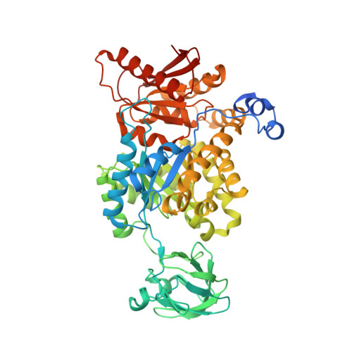 | |
UniProt & NIH Common Fund Data Resources | |||||
PHAROS: P14618 GTEx: ENSG00000067225 | |||||
Entity Groups | |||||
| Sequence Clusters | 30% Identity50% Identity70% Identity90% Identity95% Identity100% Identity | ||||
| UniProt Group | P14618 | ||||
Sequence AnnotationsExpand | |||||
Reference Sequence | |||||
| Ligands 7 Unique | |||||
|---|---|---|---|---|---|
| ID | Chains | Name / Formula / InChI Key | 2D Diagram | 3D Interactions | |
| FBP Download:Ideal Coordinates CCD File | F [auth A], Q [auth C] | 1,6-di-O-phosphono-beta-D-fructofuranose C6 H14 O12 P2 RNBGYGVWRKECFJ-ARQDHWQXSA-N |  | ||
| SO4 Download:Ideal Coordinates CCD File | M [auth B], X [auth D] | SULFATE ION O4 S QAOWNCQODCNURD-UHFFFAOYSA-L |  | ||
| PO4 Download:Ideal Coordinates CCD File | J [auth A], K [auth A], W [auth D] | PHOSPHATE ION O4 P NBIIXXVUZAFLBC-UHFFFAOYSA-K |  | ||
| GOL Download:Ideal Coordinates CCD File | AA [auth D], BA [auth D], I [auth A], P [auth B], U [auth C] | GLYCEROL C3 H8 O3 PEDCQBHIVMGVHV-UHFFFAOYSA-N |  | ||
| OXD Download:Ideal Coordinates CCD File | E [auth A], L [auth B], R [auth C], V [auth D] | OXALIC ACID C2 H2 O4 MUBZPKHOEPUJKR-UHFFFAOYSA-N |  | ||
| K Download:Ideal Coordinates CCD File | G [auth A], O [auth B], T [auth C], Z [auth D] | POTASSIUM ION K NPYPAHLBTDXSSS-UHFFFAOYSA-N |  | ||
| MG Download:Ideal Coordinates CCD File | H [auth A], N [auth B], S [auth C], Y [auth D] | MAGNESIUM ION Mg JLVVSXFLKOJNIY-UHFFFAOYSA-N |  | ||
| Length ( Å ) | Angle ( ˚ ) |
|---|---|
| a = 94.359 | α = 90 |
| b = 115.26 | β = 114.43 |
| c = 109.401 | γ = 90 |
| Software Name | Purpose |
|---|---|
| REFMAC | refinement |
| MOLREP | phasing |


