Structure of a 13-fold superhelix (almost) determined from first principles.
Schoch, G.A., Sammito, M., Millan, C., Uson, I., Rudolph, M.G.(2015) IUCrJ 2: 177-187
- PubMed: 25866655 Search on PubMedSearch on PubMed Central
- DOI: https://doi.org/10.1107/S2052252515000238
- Primary Citation Related Structures:
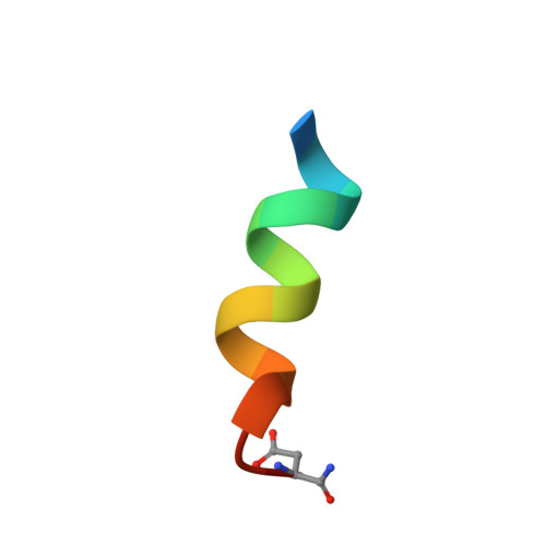
4WG0 - PubMed Abstract:
Nuclear hormone receptors are cytoplasm-based transcription factors that bind a ligand, translate to the nucleus and initiate gene transcription in complex with a co-activator such as TIF2 (transcriptional intermediary factor 2). For structural studies the co-activator is usually mimicked by a peptide of circa 13 residues, which for the largest part forms an α-helix when bound to the receptor. The aim was to co-crystallize the glucocorticoid receptor in complex with a ligand and the TIF2 co-activator peptide. The 1.82 Å resolution diffraction data obtained from the crystal could not be phased by molecular replacement using the known receptor structures. HPLC analysis of the crystals revealed the absence of the receptor and indicated that only the co-activator peptide was present. The self-rotation function displayed 13-fold rotational symmetry, which initiated an exhaustive but unsuccessful molecular-replacement approach using motifs of 13-fold symmetry such as α- and β-barrels in various geometries. The structure was ultimately determined by using a single α-helix and the software ARCIMBOLDO, which assembles fragments placed by PHASER before using them as seeds for density modification model building in SHELXE. Systematic variation of the helix length revealed upper and lower size limits for successful structure determination. A beautiful but unanticipated structure was obtained that forms superhelices with left-handed twist throughout the crystal, stabilized by ligand interactions. Together with the increasing diversity of structural elements in the Protein Data Bank the results from TIF2 confirm the potential of fragment-based molecular replacement to significantly accelerate the phasing step for native diffraction data at around 2 Å resolution.
- Molecular Design and Chemical Biology, F. Hoffmann-La Roche Ltd , Grenzacherstrasse 124, 4070 Basel, Switzerland.
Organizational Affiliation: