4WF2

Structure of E. coli BirA G142A bound to biotinol-5'-AMP


Experimental Data Snapshot

  • Method: X-RAY DIFFRACTION
  • Resolution: 2.31 Å
  • R-Value Free: 0.218 
  • R-Value Work: 0.172 
  • R-Value Observed: 0.174 

wwPDB Validation   3D Report Full Report


Ligand Structure Quality Assessment 


This is version 1.6 of the entry. See complete history


Literature

Allosteric Coupling via Distant Disorder-to-Order Transitions.

Eginton, C.Cressman, W.J.Bachas, S.Wade, H.Beckett, D.

(2015) J Mol Biol 427: 1695-1704

  • DOI: https://doi.org/10.1016/j.jmb.2015.02.021
  • Primary Citation of Related Structures:  
    4WF2

  • PubMed Abstract: 

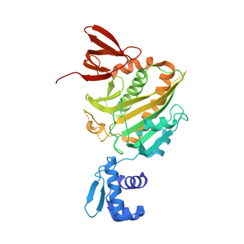
    Intrinsic disorder provides a means of maximizing allosteric coupling in proteins. However, the mechanisms by which the disorder functions in allostery remain to be elucidated. Small ligand, bio-5'-AMP, binding and dimerization of the Escherichia coli biotin repressor are allosterically coupled. Folding of a disordered loop in the allosteric effector binding site is required to realize the full coupling free energy of -4.0 ± 0.3 kcal/mol observed in the wild-type protein. Alanine substitution of a glycine residue on the dimerization surface that does not directly contribute to the dimerization interface completely abolishes this coupling. In this work, the structure of this variant, solved by X-ray crystallography, reveals a monomeric corepressor-bound protein. In the structure loops, neither of which contains the alanine substitution, on both the dimerization and effector binding surfaces that are folded in the corepressor-bound wild-type protein are disordered. The structural data combined with functional measurements indicate that allosteric coupling between ligand binding and dimerization in BirA (E. coli biotin repressor/biotin protein ligase) is achieved via reciprocal communication of disorder-to-order transitions on two distant functional surfaces.


  • Organizational Affiliation

    Department of Chemistry and Biochemistry, University of Maryland, College Park, MD 20742, USA.


Macromolecules
Find similar proteins by:  (by identity cutoff)  |  3D Structure
Entity ID: 1
MoleculeChains Sequence LengthOrganismDetailsImage
Bifunctional ligase/repressor BirA327Escherichia coli K-12Mutation(s): 1 
Gene Names: birAbioRdhbBb3973JW3941
EC: 6.3.4.15
UniProt
Find proteins for P06709 (Escherichia coli (strain K12))
Explore P06709 
Go to UniProtKB:  P06709
Entity Groups  
Sequence Clusters30% Identity50% Identity70% Identity90% Identity95% Identity100% Identity
UniProt GroupP06709
Sequence Annotations
Expand
  • Reference Sequence
Small Molecules
Ligands 1 Unique
IDChains Name / Formula / InChI Key2D Diagram3D Interactions
BTX
Query on BTX

Download Ideal Coordinates CCD File 
B [auth A]((2R,3S,4R,5R)-5-(6-AMINO-9H-PURIN-9-YL)-3,4-DIHYDROXY-TETRAHYDROFURAN-2-YL)METHYL 5-((3AS,4S,6AR)-2-OXO-HEXAHYDRO-1H-THIENO[3,4-D]IMIDAZOL-4-YL)PENTYL HYDROGEN PHOSPHATE
C20 H30 N7 O8 P S
KBOGUFFJCBPJEH-SQGSUPJISA-N
Binding Affinity Annotations 
IDSourceBinding Affinity
BTX BindingDB:  4WF2 Ki: 87 (nM) from 1 assay(s)
Kd: 1.5 (nM) from 1 assay(s)
Experimental Data & Validation

Experimental Data

  • Method: X-RAY DIFFRACTION
  • Resolution: 2.31 Å
  • R-Value Free: 0.218 
  • R-Value Work: 0.172 
  • R-Value Observed: 0.174 
  • Space Group: P 43
Unit Cell:
Length ( Å )Angle ( ˚ )
a = 46.239α = 90
b = 46.239β = 90
c = 157.107γ = 90
Software Package:
Software NamePurpose
PHENIXrefinement

Structure Validation

View Full Validation Report


Ligand Structure Quality Assessment 


Entry History & Funding Information

Deposition Data


Funding OrganizationLocationGrant Number
National Science Foundation (NSF, United States)United StatesMCB-0953430

Revision History  (Full details and data files)

  • Version 1.0: 2014-10-22
    Type: Initial release
  • Version 1.1: 2015-02-04
    Changes: Derived calculations
  • Version 1.2: 2015-03-25
    Changes: Database references
  • Version 1.3: 2015-04-08
    Changes: Database references
  • Version 1.4: 2017-09-27
    Changes: Author supporting evidence, Database references, Derived calculations, Other, Source and taxonomy, Structure summary
  • Version 1.5: 2019-11-27
    Changes: Author supporting evidence
  • Version 1.6: 2023-12-27
    Changes: Data collection, Database references, Refinement description