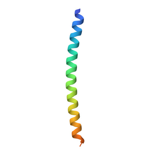
Structural Plasticity of the Coiled-Coil Domain of Rotavirus NSP4.
Sastri, N.P., Viskovska, M., Hyser, J.M., Tanner, M.R., Horton, L.B., Sankaran, B., Prasad, B.V., Estes, M.K.(2014) J Virol 88: 13602-13612
- PubMed: 25231315
- DOI: https://doi.org/10.1128/JVI.02227-14
- Primary Citation of Related Structures:
4WB4, 4WBA - PubMed Abstract:
Rotavirus (RV) nonstructural protein 4 (NSP4) is a virulence factor that disrupts cellular Ca(2+) homeostasis and plays multiple roles regulating RV replication and the pathophysiology of RV-induced diarrhea. Although its native oligomeric state is unclear, crystallographic studies of the coiled-coil domain (CCD) of NSP4 from two different strains suggest that it functions as a tetramer or a pentamer. While the CCD of simian strain SA11 NSP4 forms a tetramer that binds Ca(2+) at its core, the CCD of human strain ST3 forms a pentamer lacking the bound Ca(2+) despite the residues (E120 and Q123) that coordinate Ca(2+) binding being conserved. In these previous studies, while the tetramer crystallized at neutral pH, the pentamer crystallized at low pH, suggesting that preference for a particular oligomeric state is pH dependent and that pH could influence Ca(2+) binding. Here, we sought to examine if the CCD of NSP4 from a single RV strain can exist in two oligomeric states regulated by Ca(2+) or pH. Biochemical, biophysical, and crystallographic studies show that while the CCD of SA11 NSP4 exhibits high-affinity binding to Ca(2+) at neutral pH and forms a tetramer, it does not bind Ca(2+) at low pH and forms a pentamer, and the transition from tetramer to pentamer is reversible with pH. Mutational analysis shows that Ca(2+) binding is necessary for the tetramer formation, as an E120A mutant forms a pentamer. We propose that the structural plasticity of NSP4 regulated by pH and Ca(2+) may form a basis for its pleiotropic functions during RV replication. The nonstructural protein NSP4 of rotavirus is a multifunctional protein that plays an important role in virus replication, morphogenesis, and pathogenesis. Previous crystallography studies of the coiled-coil domain (CCD) of NSP4 from two different rotavirus strains showed two distinct oligomeric states, a Ca(2+)-bound tetrameric state and a Ca(2+)-free pentameric state. Whether NSP4 CCD from the same strain can exist in different oligomeric states and what factors might regulate its oligomeric preferences are not known. This study used a combination of biochemical, biophysical, and crystallography techniques and found that the NSP4 CCD can undergo a reversible transition from a Ca(2+)-bound tetramer to a Ca(2+)-free pentamer in response to changes in pH. From these studies, we hypothesize that this remarkable structural adaptability of the CCD forms a basis for the pleiotropic functional properties of NSP4.
- Department of Molecular Virology and Microbiology, Baylor College of Medicine, Houston, Texas, USA.
Organizational Affiliation:


