4WAA | pdb_00004waa

Crystal structure of Nix LIR-fused human LC3B_2-119


Experimental Data Snapshot

  • Method: X-RAY DIFFRACTION
  • Resolution: 2.35 Å
  • R-Value Free: 
    0.273 (Depositor), 0.277 (DCC) 
  • R-Value Work: 
    0.200 (Depositor), 0.207 (DCC) 
  • R-Value Observed: 
    0.203 (Depositor) 

Starting Model: experimental
View more details

wwPDB Validation 3D Report Full Report

Validation slider image for 4WAA

This is version 1.2 of the entry. See complete history

Literature

Phosphorylation of the mitochondrial autophagy receptor Nix enhances its interaction with LC3 proteins.

Rogov, V.V.Suzuki, H.Marinkovic, M.Lang, V.Kato, R.Kawasaki, M.Buljubasic, M.Sprung, M.Rogova, N.Wakatsuki, S.Hamacher-Brady, A.Dotsch, V.Dikic, I.Brady, N.R.Novak, I.

(2017) Sci Rep 7: 1131-1131

  • DOI: https://doi.org/10.1038/s41598-017-01258-6
  • Primary Citation Related Structures: 
    4WAA

  • PubMed Abstract: 

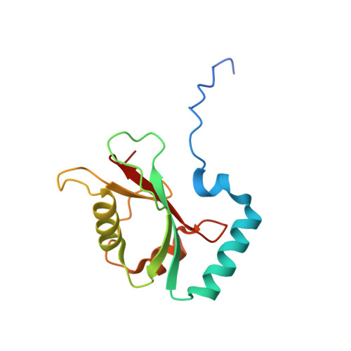
    The mitophagy receptor Nix interacts with LC3/GABARAP proteins, targeting mitochondria into autophagosomes for degradation. Here we present evidence for phosphorylation-driven regulation of the Nix:LC3B interaction. Isothermal titration calorimetry and NMR indicate a ~100 fold enhanced affinity of the serine 34/35-phosphorylated Nix LC3-interacting region (LIR) to LC3B and formation of a very rigid complex compared to the non-phosphorylated sequence. Moreover, the crystal structure of LC3B in complex with the Nix LIR peptide containing glutamic acids as phosphomimetic residues and NMR experiments revealed that LIR phosphorylation stabilizes the Nix:LC3B complex via formation of two additional hydrogen bonds between phosphorylated serines of Nix LIR and Arg11, Lys49 and Lys51 in LC3B. Substitution of Lys51 to Ala in LC3B abrogates binding of a phosphomimetic Nix mutant. Functionally, serine 34/35 phosphorylation enhances autophagosome recruitment to mitochondria in HeLa cells. Together, this study provides cellular, biochemical and biophysical evidence that phosphorylation of the LIR domain of Nix enhances mitophagy receptor engagement.


  • Organizational Affiliation
    • Institute of Biophysical Chemistry and Center for Biomolecular Magnetic Resonance, Goethe University, D-60438, Frankfurt, Germany.

Macromolecule Content 

  • Total Structure Weight: 31.17 kDa 
  • Atom Count: 2,194 
  • Modeled Residue Count: 251 
  • Deposited Residue Count: 268 
  • Unique protein chains: 1

Macromolecules

Find similar proteins by:|  3D Structure
Entity ID: 1
MoleculeChains  Sequence LengthOrganismDetailsImage
Microtubule-associated proteins 1A/1B light chain 3B
A, B
134Homo sapiensMutation(s): 0 
Gene Names: MAP1LC3BMAP1ALC3
UniProt & NIH Common Fund Data Resources
Find proteins for Q9GZQ8 (Homo sapiens)
Explore Q9GZQ8 
Go to UniProtKB:  Q9GZQ8
PHAROS:  Q9GZQ8
GTEx:  ENSG00000140941 
Entity Groups
Sequence Clusters30% Identity50% Identity70% Identity90% Identity95% Identity100% Identity
UniProt GroupQ9GZQ8
Sequence Annotations
Expand
Reference Sequence

Experimental Data & Validation

Experimental Data

  • Method: X-RAY DIFFRACTION
  • Resolution: 2.35 Å
  • R-Value Free:  0.273 (Depositor), 0.277 (DCC) 
  • R-Value Work:  0.200 (Depositor), 0.207 (DCC) 
  • R-Value Observed: 0.203 (Depositor) 
Space Group: P 43 21 2
Unit Cell:
Length ( Å )Angle ( ˚ )
a = 67.385α = 90
b = 67.385β = 90
c = 116.875γ = 90
Software Package:
Software NamePurpose
REFMACrefinement
iMOSFLMdata reduction
SCALAdata scaling
MOLREPphasing

Structure Validation

View Full Validation Report


Entry History 

Deposition Data

Revision History  (Full details and data files)

  • Version 1.0: 2015-09-09
    Type: Initial release
  • Version 1.1: 2018-06-20
    Changes: Data collection, Database references
  • Version 1.2: 2024-01-10
    Changes: Data collection, Database references, Refinement description