4W9H | pdb_00004w9h

pVHL:EloB:EloC in complex with (2S,4R)-1-((S)-2-acetamido-3,3-dimethylbutanoyl)-4-hydroxy-N-(4-(4-methylthiazol-5-yl)benzyl)pyrrolidine-2-carboxamide (ligand 7)


Experimental Data Snapshot

  • Method: X-RAY DIFFRACTION
  • Resolution: 2.10 Å
  • R-Value Free: 
    0.253 (Depositor), 0.259 (DCC) 
  • R-Value Work: 
    0.210 (Depositor), 0.217 (DCC) 
  • R-Value Observed: 
    0.212 (Depositor) 

Starting Model: experimental
View more details

wwPDB Validation 3D Report Full Report

Validation slider image for 4W9H

Ligand Structure Quality Assessment 


This is version 1.3 of the entry. See complete history

Literature

Structure-Guided Design and Optimization of Small Molecules Targeting the Protein-Protein Interaction between the von Hippel-Lindau (VHL) E3 Ubiquitin Ligase and the Hypoxia Inducible Factor (HIF) Alpha Subunit with in Vitro Nanomolar Affinities.

Galdeano, C.Gadd, M.S.Soares, P.Scaffidi, S.Van Molle, I.Birced, I.Hewitt, S.Dias, D.M.Ciulli, A.

(2014) J Med Chem 57: 8657-8663

  • DOI: https://doi.org/10.1021/jm5011258
  • Primary Citation Related Structures: 
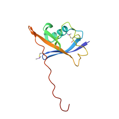
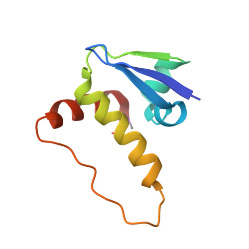
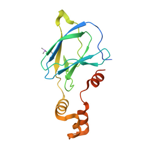
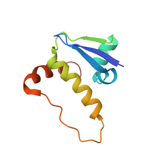
    4W9C, 4W9D, 4W9E, 4W9F, 4W9G, 4W9H, 4W9I, 4W9J, 4W9K, 4W9L

  • PubMed Abstract: 

    E3 ubiquitin ligases are attractive targets in the ubiquitin-proteasome system, however, the development of small-molecule ligands has been rewarded with limited success. The von Hippel-Lindau protein (pVHL) is the substrate recognition subunit of the VHL E3 ligase that targets HIF-1α for degradation. We recently reported inhibitors of the pVHL:HIF-1α interaction, however they exhibited moderate potency. Herein, we report the design and optimization, guided by X-ray crystal structures, of a ligand series with nanomolar binding affinities.


  • Organizational Affiliation
    • Division of Biological Chemistry and Drug Discovery, College of Life Sciences, University of Dundee , Dow Street, Dundee, DD1 5EH, Scotland, U.K.

Macromolecule Content 

  • Total Structure Weight: 169.05 kDa 
  • Atom Count: 11,271 
  • Modeled Residue Count: 1,332 
  • Deposited Residue Count: 1,452 
  • Unique protein chains: 4

Macromolecules

Find similar proteins by:|  3D Structure
Entity ID: 1
MoleculeChains  Sequence LengthOrganismDetailsImage
Transcription elongation factor B polypeptide 2
A, D, G, J
104Homo sapiensMutation(s): 0 
Gene Names: TCEB2
UniProt & NIH Common Fund Data Resources
Find proteins for Q15370 (Homo sapiens)
Explore Q15370 
Go to UniProtKB:  Q15370
PHAROS:  Q15370
GTEx:  ENSG00000103363 
Entity Groups
Sequence Clusters30% Identity50% Identity70% Identity90% Identity95% Identity100% Identity
UniProt GroupQ15370
Sequence Annotations
Expand
Reference Sequence
Find similar proteins by:|  3D Structure
Entity ID: 2
MoleculeChains  Sequence LengthOrganismDetailsImage
Transcription elongation factor B polypeptide 1
B, E
97Homo sapiensMutation(s): 0 
Gene Names: TCEB1
UniProt & NIH Common Fund Data Resources
Find proteins for Q15369 (Homo sapiens)
Explore Q15369 
Go to UniProtKB:  Q15369
PHAROS:  Q15369
GTEx:  ENSG00000154582 
Entity Groups
Sequence Clusters30% Identity50% Identity70% Identity90% Identity95% Identity100% Identity
UniProt GroupQ15369
Sequence Annotations
Expand
Reference Sequence
Find similar proteins by:|  3D Structure
Entity ID: 3
MoleculeChains  Sequence LengthOrganismDetailsImage
Von Hippel-Lindau disease tumor suppressor
C, F, I, L
162Homo sapiensMutation(s): 0 
Gene Names: VHL
UniProt & NIH Common Fund Data Resources
Find proteins for P40337 (Homo sapiens)
Explore P40337 
Go to UniProtKB:  P40337
PHAROS:  P40337
GTEx:  ENSG00000134086 
Entity Groups
Sequence Clusters30% Identity50% Identity70% Identity90% Identity95% Identity100% Identity
UniProt GroupP40337
Sequence Annotations
Expand
Reference Sequence
Find similar proteins by:|  3D Structure
Entity ID: 4
MoleculeChains  Sequence LengthOrganismDetailsImage
Transcription elongation factor B polypeptide 1
H, K
97Homo sapiensMutation(s): 0 
Gene Names: TCEB1
UniProt & NIH Common Fund Data Resources
Find proteins for Q15369 (Homo sapiens)
Explore Q15369 
Go to UniProtKB:  Q15369
PHAROS:  Q15369
GTEx:  ENSG00000154582 
Entity Groups
Sequence Clusters30% Identity50% Identity70% Identity90% Identity95% Identity100% Identity
UniProt GroupQ15369
Sequence Annotations
Expand
Reference Sequence

Experimental Data & Validation

Experimental Data

  • Method: X-RAY DIFFRACTION
  • Resolution: 2.10 Å
  • R-Value Free:  0.253 (Depositor), 0.259 (DCC) 
  • R-Value Work:  0.210 (Depositor), 0.217 (DCC) 
  • R-Value Observed: 0.212 (Depositor) 
Space Group: P 41 2 2
Unit Cell:
Length ( Å )Angle ( ˚ )
a = 92.98α = 90
b = 92.98β = 90
c = 364.372γ = 90
Software Package:
Software NamePurpose
SCALAdata scaling
REFMACrefinement
PDB_EXTRACTdata extraction
XDSdata reduction
XSCALEdata scaling
XSCALEdata reduction

Structure Validation

View Full Validation Report


Ligand Structure Quality Assessment 


Entry History 

& Funding Information

Deposition Data


Funding OrganizationLocationGrant Number
European Research CouncilERC-2012-StG-311460
Biotechnology and Biological Sciences Research CouncilUnited KingdomBBSRC BB/G023123/1
Marie Sklodowska-Curie ActionsEC PIEF-GA-2012-328030

Revision History  (Full details and data files)

  • Version 1.0: 2014-09-10
    Type: Initial release
  • Version 1.1: 2014-11-05
    Changes: Database references
  • Version 1.2: 2024-01-10
    Changes: Data collection, Database references, Refinement description
  • Version 1.3: 2024-10-23
    Changes: Structure summary