4UXX | pdb_00004uxx

Structure of delta4-DgkA with AMPPCP in 9.9 MAG


Experimental Data Snapshot

  • Method: X-RAY DIFFRACTION
  • Resolution: 2.70 Å
  • R-Value Free: 
    0.258 (Depositor), 0.263 (DCC) 
  • R-Value Work: 
    0.219 (Depositor), 0.235 (DCC) 
  • R-Value Observed: 
    0.221 (Depositor) 

Starting Model: experimental
View more details

wwPDB Validation 3D Report Full Report

Validation slider image for 4UXX

Ligand Structure Quality Assessment 


This is version 2.2 of the entry. See complete history

Literature

Ternary Structure Reveals Mechanism of a Membrane Diacylglycerol Kinase.

Li, D.Stansfeld, P.J.Sansom, M.S.P.Keogh, A.Vogeley, L.Howe, N.Lyons, J.A.Aragao, D.Fromme, P.Fromme, R.Basu, S.Grotjohann, I.Kupitz, C.Rendek, K.Weierstall, U.Zatsepin, N.A.Cherezov, V.Liu, W.Bandaru, S.English, N.J.Gati, C.Barty, A.Yefanov, O.Chapman, H.N.Diederichs, K.Messerschmidt, M.Boutet, S.Williams, G.J.Marvin Seibert, M.Caffrey, M.

(2015) Nat Commun 6: 10140

  • DOI: https://doi.org/10.1038/ncomms10140
  • Primary Citation Related Structures: 
    4UXW, 4UXX, 4UXZ, 4UYO

  • PubMed Abstract: 

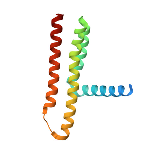
    Diacylglycerol kinase catalyses the ATP-dependent conversion of diacylglycerol to phosphatidic acid in the plasma membrane of Escherichia coli. The small size of this integral membrane trimer, which has 121 residues per subunit, means that available protein must be used economically to craft three catalytic and substrate-binding sites centred about the membrane/cytosol interface. How nature has accomplished this extraordinary feat is revealed here in a crystal structure of the kinase captured as a ternary complex with bound lipid substrate and an ATP analogue. Residues, identified as essential for activity by mutagenesis, decorate the active site and are rationalized by the ternary structure. The γ-phosphate of the ATP analogue is positioned for direct transfer to the primary hydroxyl of the lipid whose acyl chain is in the membrane. A catalytic mechanism for this unique enzyme is proposed. The active site architecture shows clear evidence of having arisen by convergent evolution.


  • Organizational Affiliation
    • School of Medicine and School of Biochemistry and Immunology, Trinity College Dublin, Dublin 2, Ireland.

Macromolecule Content 

  • Total Structure Weight: 45.9 kDa 
  • Atom Count: 2,804 
  • Modeled Residue Count: 336 
  • Deposited Residue Count: 390 
  • Unique protein chains: 1

Macromolecules

Find similar proteins by:|  3D Structure
Entity ID: 1
MoleculeChains  Sequence LengthOrganismDetailsImage
DIACYLGLYCEROL KINASE
A, B, C
130Escherichia coli K-12Mutation(s): 4 
EC: 2.7.1.107
Membrane Entity: Yes 
UniProt
Find proteins for P0ABN1 (Escherichia coli (strain K12))
Explore P0ABN1 
Go to UniProtKB:  P0ABN1
Entity Groups
Sequence Clusters30% Identity50% Identity70% Identity90% Identity95% Identity100% Identity
UniProt GroupP0ABN1
Sequence Annotations
Expand
Reference Sequence

Small Molecules

Ligands 7 Unique
IDChains Name / Formula / InChI Key2D Diagram3D Interactions
ACP

Query on ACP


Download:Ideal Coordinates CCD File
Q [auth C]PHOSPHOMETHYLPHOSPHONIC ACID ADENYLATE ESTER
C11 H18 N5 O12 P3
UFZTZBNSLXELAL-IOSLPCCCSA-N
OLC

Query on OLC


Download:Ideal Coordinates CCD File
G [auth A]
H [auth A]
K [auth B]
L [auth B]
R [auth C]
G [auth A],
H [auth A],
K [auth B],
L [auth B],
R [auth C],
S [auth C]
(2R)-2,3-dihydroxypropyl (9Z)-octadec-9-enoate
C21 H40 O4
RZRNAYUHWVFMIP-GDCKJWNLSA-N
MPD

Query on MPD


Download:Ideal Coordinates CCD File
J [auth B](4S)-2-METHYL-2,4-PENTANEDIOL
C6 H14 O2
SVTBMSDMJJWYQN-YFKPBYRVSA-N
ZN

Query on ZN


Download:Ideal Coordinates CCD File
O [auth C],
P [auth C]
ZINC ION
Zn
PTFCDOFLOPIGGS-UHFFFAOYSA-N
ACT

Query on ACT


Download:Ideal Coordinates CCD File
F [auth A],
I [auth A],
M [auth B]
ACETATE ION
C2 H3 O2
QTBSBXVTEAMEQO-UHFFFAOYSA-M
CL

Query on CL


Download:Ideal Coordinates CCD File
D [auth A]CHLORIDE ION
Cl
VEXZGXHMUGYJMC-UHFFFAOYSA-M
NA

Query on NA


Download:Ideal Coordinates CCD File
E [auth A],
N [auth B],
T [auth C]
SODIUM ION
Na
FKNQFGJONOIPTF-UHFFFAOYSA-N

Experimental Data & Validation

Experimental Data

  • Method: X-RAY DIFFRACTION
  • Resolution: 2.70 Å
  • R-Value Free:  0.258 (Depositor), 0.263 (DCC) 
  • R-Value Work:  0.219 (Depositor), 0.235 (DCC) 
  • R-Value Observed: 0.221 (Depositor) 
Space Group: P 31 2 1
Unit Cell:
Length ( Å )Angle ( ˚ )
a = 72.94α = 90
b = 72.94β = 90
c = 195.76γ = 120
Software Package:
Software NamePurpose
PHENIXrefinement
XDSdata reduction
XSCALEdata scaling
PHASERphasing

Structure Validation

View Full Validation Report


Ligand Structure Quality Assessment 


Entry History 

Deposition Data

Revision History  (Full details and data files)

  • Version 1.0: 2015-09-30
    Type: Initial release
  • Version 1.1: 2016-01-13
    Changes: Database references
  • Version 2.0: 2019-03-06
    Changes: Atomic model, Data collection, Experimental preparation, Other
  • Version 2.1: 2019-04-03
    Changes: Data collection, Experimental preparation
  • Version 2.2: 2024-01-10
    Changes: Data collection, Database references, Derived calculations, Other, Refinement description