4UWM | pdb_00004uwm

Type II Baeyer-Villiger monooxygenase.The oxygenating constituent of 3,6-diketocamphane monooxygenase from CAM plasmid of Pseudomonas putida in complex with FMN.


Experimental Data Snapshot

  • Method: X-RAY DIFFRACTION
  • Resolution: 1.90 Å
  • R-Value Free: 
    0.221 (Depositor), 0.221 (DCC) 
  • R-Value Work: 
    0.183 (Depositor), 0.183 (DCC) 
  • R-Value Observed: 
    0.185 (Depositor) 

Starting Model: experimental
View more details

wwPDB Validation 3D Report Full Report

Validation slider image for 4UWM

Ligand Structure Quality Assessment 


This is version 1.2 of the entry. See complete history

Literature

The Oxygenating Constituent of 3,6-Diketocamphane Monooxygenase from the Cam Plasmid of Pseudomonas Putida: The First Crystal Structure of a Type II Baeyer-Villiger Monooxygenase.

Isupov, M.N.Schroder, E.Gibson, R.P.Beecher, J.Donadio, G.Saneei, V.Dcunha, S.A.Mcghie, E.J.Sayer, C.Davenport, C.F.Lau, P.C.Hasegawa, Y.Iwaki, H.Kadow, M.Balke, K.Bornscheuer, U.T.Bourenkov, G.Littlechild, J.A.

(2015) Acta Crystallogr D Biol Crystallogr 71: 2344

  • DOI: https://doi.org/10.1107/S1399004715017939
  • Primary Citation Related Structures: 
    4UWM, 5AEC

  • PubMed Abstract: 

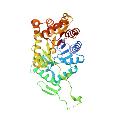
    The three-dimensional structures of the native enzyme and the FMN complex of the overexpressed form of the oxygenating component of the type II Baeyer-Villiger 3,6-diketocamphane monooxygenase have been determined to 1.9 Å resolution. The structure of this dimeric FMN-dependent enzyme, which is encoded on the large CAM plasmid of Pseudomonas putida, has been solved by a combination of multiple anomalous dispersion from a bromine crystal soak and molecular replacement using a bacterial luciferase model. The orientation of the isoalloxazine ring of the FMN cofactor in the active site of this TIM-barrel fold enzyme differs significantly from that previously observed in enzymes of the bacterial luciferase-like superfamily. The Ala77 residue is in a cis conformation and forms a β-bulge at the C-terminus of β-strand 3, which is a feature observed in many proteins of this superfamily.


  • Organizational Affiliation
    • The Henry Wellcome Building for Biocatalysis, Biosciences, College of Life and Environmental Sciences, University of Exeter, Stocker Road, Exeter EX4 4QD, England.

Macromolecule Content 

  • Total Structure Weight: 86.8 kDa 
  • Atom Count: 7,027 
  • Modeled Residue Count: 724 
  • Deposited Residue Count: 756 
  • Unique protein chains: 1

Macromolecules

Find similar proteins by:|  3D Structure
Entity ID: 1
MoleculeChains  Sequence LengthOrganismDetailsImage
3,6-DIKETOCAMPHANE 1,6 MONOOXYGENASE
A, B
378Pseudomonas putidaMutation(s): 0 
EC: 1.14.13 (PDB Primary Data), 1.14.14.155 (UniProt)
UniProt
Find proteins for D7UER1 (Pseudomonas putida)
Explore D7UER1 
Go to UniProtKB:  D7UER1
Entity Groups
Sequence Clusters30% Identity50% Identity70% Identity90% Identity95% Identity100% Identity
UniProt GroupD7UER1
Sequence Annotations
Expand
Reference Sequence

Small Molecules

Ligands 4 Unique
IDChains Name / Formula / InChI Key2D Diagram3D Interactions
FMN

Query on FMN


Download:Ideal Coordinates CCD File
C [auth A],
H [auth B]
FLAVIN MONONUCLEOTIDE
C17 H21 N4 O9 P
FVTCRASFADXXNN-SCRDCRAPSA-N
PG4

Query on PG4


Download:Ideal Coordinates CCD File
D [auth A],
I [auth B],
L [auth B]
TETRAETHYLENE GLYCOL
C8 H18 O5
UWHCKJMYHZGTIT-UHFFFAOYSA-N
PGE

Query on PGE


Download:Ideal Coordinates CCD File
G [auth A]TRIETHYLENE GLYCOL
C6 H14 O4
ZIBGPFATKBEMQZ-UHFFFAOYSA-N
PEG

Query on PEG


Download:Ideal Coordinates CCD File
E [auth A],
F [auth A],
J [auth B],
K [auth B]
DI(HYDROXYETHYL)ETHER
C4 H10 O3
MTHSVFCYNBDYFN-UHFFFAOYSA-N

Experimental Data & Validation

Experimental Data

  • Method: X-RAY DIFFRACTION
  • Resolution: 1.90 Å
  • R-Value Free:  0.221 (Depositor), 0.221 (DCC) 
  • R-Value Work:  0.183 (Depositor), 0.183 (DCC) 
  • R-Value Observed: 0.185 (Depositor) 
Space Group: P 21 21 21
Unit Cell:
Length ( Å )Angle ( ˚ )
a = 72.79α = 90
b = 82.84β = 90
c = 149.85γ = 90
Software Package:
Software NamePurpose
REFMACrefinement
XDSdata reduction
xia2data reduction
Aimlessdata scaling
MOLREPphasing

Structure Validation

View Full Validation Report


Ligand Structure Quality Assessment 


Entry History 

Revision History  (Full details and data files)

  • Version 1.0: 2015-08-26
    Type: Initial release
  • Version 1.1: 2015-11-11
    Changes: Database references
  • Version 1.2: 2024-01-10
    Changes: Data collection, Database references, Derived calculations, Other, Refinement description