4UW4

Human galectin-7 in complex with a galactose based dendron D2-1.


Experimental Data Snapshot

  • Method: X-RAY DIFFRACTION
  • Resolution: 1.77 Å
  • R-Value Free: 0.235 
  • R-Value Work: 0.182 
  • R-Value Observed: 0.184 

wwPDB Validation   3D Report Full Report


Ligand Structure Quality Assessment 


This is version 1.3 of the entry. See complete history


Literature

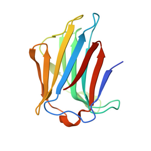
Structural Basis of Multivalent Galactose-Based Dendrimer Recognition by Human Galectin-7.

Ramaswamy, S.Haj Sleiman, M.Masuyer, G.Arbez-Gindre, C.Micha-Screttas, M.Calogeropoulou, T.Steele, B.R.Acharya, K.R.

(2015) FEBS J 282: 372

  • DOI: https://doi.org/10.1111/febs.13140
  • Primary Citation of Related Structures:  
    4UW3, 4UW4, 4UW5, 4UW6

  • PubMed Abstract: 

    Galectins are evolutionarily conserved and ubiquitously present animal lectins with a high affinity for β-galactose-containing oligosaccharides. To date, 15 mammalian galectins have been identified. Their involvement in cell-cell and cell-matrix interactions has highlighted their importance in signal transduction and other intracellular processes. Human galectin-7 (hGal-7) is a 15 kDa proto type galectin that forms a dimer in solution and its involvement in the stimulation and development of tumour growth has been reported. Previously, we reported the crystal structure of hGal-7 and its complex with galactose and lactose which provided insight into its molecular recognition and detailed interactions. Here, we present newly obtained high-resolution structural data on carbohydrate-based dendrons in complex with hGal-7. Our crystallographic data reveal how multivalent ligands interact with and form cross-links with these galectin molecules. Understanding how these dendrimeric compounds interact with hGal-7 would help in the design of new tools to investigate the recognition of carbohydrates by lectins.


  • Organizational Affiliation

    Department of Biology and Biochemistry, University of Bath, UK.


Macromolecules
Find similar proteins by:  (by identity cutoff)  |  3D Structure
Entity ID: 1
MoleculeChains Sequence LengthOrganismDetailsImage
GALECTIN-7
A, B
136Homo sapiensMutation(s): 0 
UniProt & NIH Common Fund Data Resources
Find proteins for P47929 (Homo sapiens)
Explore P47929 
Go to UniProtKB:  P47929
PHAROS:  P47929
Entity Groups  
Sequence Clusters30% Identity50% Identity70% Identity90% Identity95% Identity100% Identity
UniProt GroupP47929
Sequence Annotations
Expand
  • Reference Sequence
Small Molecules
Ligands 1 Unique
IDChains Name / Formula / InChI Key2D Diagram3D Interactions
4S0
Query on 4S0

Download Ideal Coordinates CCD File 
C [auth B]DENDRON D2-1
C49 H80 N10 O33
SJELSRWZDPHJDW-KWHDUCOQSA-N
Experimental Data & Validation

Experimental Data

  • Method: X-RAY DIFFRACTION
  • Resolution: 1.77 Å
  • R-Value Free: 0.235 
  • R-Value Work: 0.182 
  • R-Value Observed: 0.184 
  • Space Group: P 21 21 21
Unit Cell:
Length ( Å )Angle ( ˚ )
a = 38.95α = 90
b = 58.49β = 90
c = 115γ = 90
Software Package:
Software NamePurpose
PHENIXrefinement
MOSFLMdata reduction
Aimlessdata scaling
PHASERphasing

Structure Validation

View Full Validation Report


Ligand Structure Quality Assessment 


Entry History 

Deposition Data

Revision History  (Full details and data files)

  • Version 1.0: 2014-11-12
    Type: Initial release
  • Version 1.1: 2015-08-12
    Changes: Database references
  • Version 1.2: 2019-09-25
    Changes: Data collection, Experimental preparation, Other
  • Version 1.3: 2024-01-10
    Changes: Data collection, Database references, Derived calculations, Refinement description