A Flexible Extension of the Drosophila Ultrabithorax Homeodomain Defines a Novel Hox/Pbc Interaction Mode.
Foos, N., Maurel-Zaffran, C., Mate, M.J., Vincentelli, R., Hainaut, M., Berenger, H., Pradel, J., Saurin, A.J., Ortiz-Lombardia, M., Graba, Y.(2015) Structure 23: 270
- PubMed: 25651060 Search on PubMed
- DOI: https://doi.org/10.1016/j.str.2014.12.011
- Primary Citation Related Structures:
4CYC, 4UUS, 4UUT - PubMed Abstract:
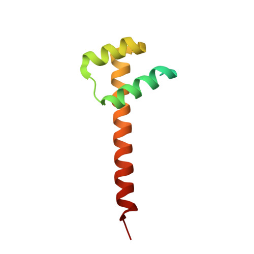
The patterning function of Hox proteins relies on assembling protein complexes with PBC proteins, which often involves a protein motif found in most Hox proteins, the so-called Hexapeptide (HX). Hox/PBC complexes likely gained functional diversity by acquiring additional modes of interaction. Here, we structurally characterize the first HX alternative interaction mode based on the paralogue-specific UbdA motif and further functionally validate structure-based predictions. The UbdA motif folds as a flexible extension of the homeodomain recognition helix and defines Hox/PBC contacts that occur, compared with those mediated by the HX motif, on the opposing side of the DNA double helix. This provides a new molecular facet to Hox/PBC complex assembly and suggests possible mechanisms for the diversification of Hox protein function.
- Centre National de la Recherche Scientifique, Aix-Marseille Université, CNRS UMR 7257, AFMB, 163 Avenue de Luminy, 13288 Marseille, France.
Organizational Affiliation: