Genomic Rearrangements and Functional Diversification of Leca and Lecb Lectin-Coding Regions Impacting the Efficacy of Glycomimetics Directed Against Pseudomonas Aeruginosa.
Boukerb, A.M., Decor, A., Ribun, S., Tabaroni, R., Rousset, A., Commin, L., Buff, S., Doleans-Jordheim, A., Vidal, S., Varrot, A., Imberty, A., Cournoyer, B.(2016) Front Microbiol 7: 811
- PubMed: 27303392 Search on PubMedSearch on PubMed Central
- DOI: https://doi.org/10.3389/fmicb.2016.00811
- Primary Citation Related Structures:
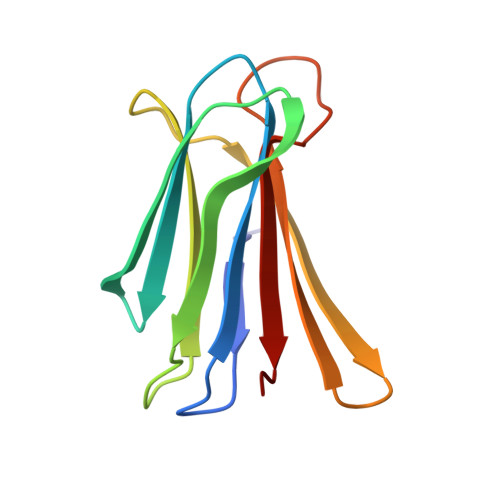
4UT5 - PubMed Abstract:
LecA and LecB tetrameric lectins take part in oligosaccharide-mediated adhesion-processes of Pseudomonas aeruginosa. Glycomimetics have been designed to block these interactions. The great versatility of P. aeruginosa suggests that the range of application of these glycomimetics could be restricted to genotypes with particular lectin types. The likelihood of having genomic and genetic changes impacting LecA and LecB interactions with glycomimetics such as galactosylated and fucosylated calix[4]arene was investigated over a collection of strains from the main clades of P. aeruginosa. Lectin types were defined, and their ligand specificities were inferred. These analyses showed a loss of lecA among the PA7 clade. Genomic changes impacting lec loci were thus assessed using strains of this clade, and by making comparisons with the PAO1 genome. The lecA regions were found challenged by phage attacks and PAGI-2 (genomic island) integrations. A prophage was linked to the loss of lecA. The lecB regions were found less impacted by such rearrangements but greater lecB than lecA genetic divergences were recorded. Sixteen combinations of LecA and LecB types were observed. Amino acid variations were mapped on PAO1 crystal structures. Most significant changes were observed on LecBPA7, and found close to the fucose binding site. Glycan array analyses were performed with purified LecBPA7. LecBPA7 was found less specific for fucosylated oligosaccharides than LecBPAO1, with a preference for H type 2 rather than type 1, and Lewis(a) rather than Lewis(x). Comparison of the crystal structures of LecBPA7 and LecBPAO1 in complex with Lewis(a) showed these changes in specificity to have resulted from a modification of the water network between the lectin, galactose and GlcNAc residues. Incidence of these modifications on the interactions with calix[4]arene glycomimetics at the cell level was investigated. An aggregation test was used to establish the efficacy of these ligands. Great variations in the responses were observed. Glycomimetics directed against LecB yielded the highest numbers of aggregates for strains from all clades. The use of a PAO1ΔlecB strain confirmed a role of LecB in this aggregation phenotype. Fucosylated calix[4]arene showed the greatest potential for a use in the prevention of P. aeruginosa infections.
- Equipes de Recherche, Bactéries Pathogènes Opportunistes et Environnement, Centre de Ressources Biologiques - Environnement Microbiologie Lyon, UMR Centre National de la Recherche Scientifique 5557 Ecologie Microbienne, Université Lyon 1 and VetAgro Sup Lyon, France.
Organizational Affiliation: