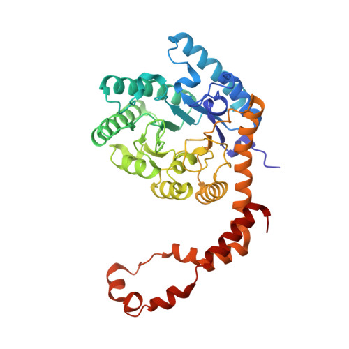
4US6

New Crystal Form of Glucose Isomerase Grown in Short Peptide Supramolecular Hydrogels


Experimental Data Snapshot

  • Method: X-RAY DIFFRACTION
  • Resolution: 1.20 Å
  • R-Value Free: 0.132 
  • R-Value Work: 0.106 
  • R-Value Observed: 0.108 

wwPDB Validation   3D Report Full Report


This is version 1.2 of the entry. See complete history


Literature

Influence of the Chirality of Short Peptide Supramolecular Hydrogels in Protein Crystallogenesis.

Conejero-Muriel, M.Gavira, J.A.Pineda-Molina, E.Belsom, A.Bradley, M.Moral, M.Garcia-Lopez Duran, J.D.D.Luque Gonzalez, A.Diaz-Mochon, J.J.Contreras-Montoya, R.Martinez-Peragon, A.Cuerva, J.M.Alvarez De Cienfuegos, L.

(2015) Chem Commun (Camb) 51: 3862

  • DOI: https://doi.org/10.1039/c4cc09024a
  • Primary Citation of Related Structures:  
    4US6

  • PubMed Abstract: 

    For the first time the influence of the chirality of the gel fibers in protein crystallogenesis has been studied. Enantiomeric hydrogels 1 and 2 were tested with model proteins lysozyme and glucose isomerase and a formamidase extracted from B. cereus. Crystallization behaviour and crystal quality of these proteins in both hydrogels are presented and compared.


  • Organizational Affiliation

    Laboratorio de Estudios Cristalográficos, Instituto Andaluz de Ciencias de la Tierra (CSIC-UGR), Av. de las Palmeras 4, 18100 Armilla, Granada, Spain. jgavira@iact.ugr-csic.es.


Macromolecules
Find similar proteins by:  (by identity cutoff)  |  3D Structure
Entity ID: 1
MoleculeChains Sequence LengthOrganismDetailsImage
XYLOSE ISOMERASE
A, B
388Streptomyces rubiginosusMutation(s): 0 
EC: 5.3.1.5
UniProt
Find proteins for P24300 (Streptomyces rubiginosus)
Explore P24300 
Go to UniProtKB:  P24300
Entity Groups  
Sequence Clusters30% Identity50% Identity70% Identity90% Identity95% Identity100% Identity
UniProt GroupP24300
Sequence Annotations
Expand
  • Reference Sequence
Small Molecules
Ligands 4 Unique
IDChains Name / Formula / InChI Key2D Diagram3D Interactions
GOL
Query on GOL

Download Ideal Coordinates CCD File 
E [auth A]
F [auth A]
G [auth A]
L [auth B]
M [auth B]
E [auth A],
F [auth A],
G [auth A],
L [auth B],
M [auth B],
N [auth B]
GLYCEROL
C3 H8 O3
PEDCQBHIVMGVHV-UHFFFAOYSA-N
CA
Query on CA

Download Ideal Coordinates CCD File 
C [auth A],
J [auth B]
CALCIUM ION
Ca
BHPQYMZQTOCNFJ-UHFFFAOYSA-N
MG
Query on MG

Download Ideal Coordinates CCD File 
D [auth A],
I [auth A],
K [auth B]
MAGNESIUM ION
Mg
JLVVSXFLKOJNIY-UHFFFAOYSA-N
NA
Query on NA

Download Ideal Coordinates CCD File 
H [auth A],
O [auth B]
SODIUM ION
Na
FKNQFGJONOIPTF-UHFFFAOYSA-N
Experimental Data & Validation

Experimental Data

  • Method: X-RAY DIFFRACTION
  • Resolution: 1.20 Å
  • R-Value Free: 0.132 
  • R-Value Work: 0.106 
  • R-Value Observed: 0.108 
  • Space Group: P 21 21 2
Unit Cell:
Length ( Å )Angle ( ˚ )
a = 85.999α = 90
b = 93.677β = 90
c = 99.215γ = 90
Software Package:
Software NamePurpose
REFMACrefinement
XDSdata reduction
SCALAdata scaling
MOLREPphasing

Structure Validation

View Full Validation Report


Entry History 

Deposition Data

Revision History  (Full details and data files)

  • Version 1.0: 2015-05-13
    Type: Initial release
  • Version 1.1: 2017-09-13
    Changes: Data collection
  • Version 1.2: 2024-01-10
    Changes: Data collection, Database references, Derived calculations, Other, Refinement description