4UMO | pdb_00004umo

Crystal Structure of the Kv7.1 proximal C-terminal Domain in Complex with Calmodulin


Experimental Data Snapshot

  • Method: X-RAY DIFFRACTION
  • Resolution: 3.00 Å
  • R-Value Free: 
    0.247 (Depositor), 0.252 (DCC) 
  • R-Value Work: 
    0.213 (Depositor), 0.224 (DCC) 
  • R-Value Observed: 
    0.215 (Depositor) 

wwPDB Validation 3D Report Full Report

Validation slider image for 4UMO

This is version 1.3 of the entry. See complete history

Literature

Structural Basis of a Kv7.1 Potassium Channel Gating Module: Studies of the Intracellular C-Terminal Domain in Complex with Calmodulin

Sachyani, D.Dvir, M.Strulovich, R.Tria, G.Tobelaim, W.Peretz, A.Pongs, O.Svergun, D.Attali, B.Hirsch, J.A.

(2014) Structure 22: 1582

  • DOI: https://doi.org/10.1016/j.str.2014.07.016
  • Primary Citation Related Structures: 
    4UMO, 4V0C

  • PubMed Abstract: 

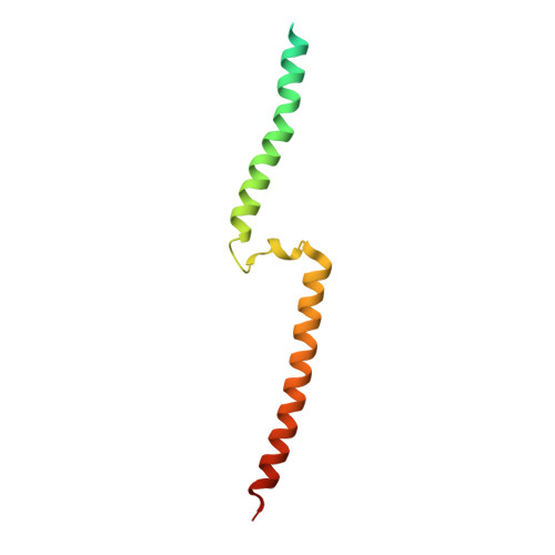
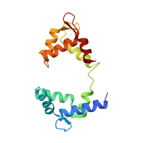
    Kv7 channels tune neuronal and cardiomyocyte excitability. In addition to the channel membrane domain, they also have a unique intracellular C-terminal (CT) domain, bound constitutively to calmodulin (CaM). This CT domain regulates gating and tetramerization. We investigated the structure of the membrane proximal CT module in complex with CaM by X-ray crystallography. The results show how the CaM intimately hugs a two-helical bundle, explaining many channelopathic mutations. Structure-based mutagenesis of this module in the context of concatemeric tetramer channels and functional analysis along with in vitro data lead us to propose that one CaM binds to one individual protomer, without crosslinking subunits and that this configuration is required for proper channel expression and function. Molecular modeling of the CT/CaM complex in conjunction with small-angle X-ray scattering suggests that the membrane proximal region, having a rigid lever arm, is a critical gating regulator.

Macromolecule Content 

  • Total Structure Weight: 60.96 kDa 
  • Atom Count: 3,518 
  • Modeled Residue Count: 459 
  • Deposited Residue Count: 522 
  • Unique protein chains: 2

Macromolecules

Find similar proteins by:|  3D Structure
Entity ID: 1
MoleculeChains  Sequence LengthOrganismDetailsImage
POTASSIUM VOLTAGE-GATED CHANNEL SUBFAMILY KQT MEMBER 1
A, B
112Homo sapiensMutation(s): 2 
UniProt & NIH Common Fund Data Resources
Find proteins for P51787 (Homo sapiens)
Explore P51787 
Go to UniProtKB:  P51787
PHAROS:  P51787
GTEx:  ENSG00000053918 
Entity Groups
Sequence Clusters30% Identity50% Identity70% Identity90% Identity95% Identity100% Identity
UniProt GroupP51787
Sequence Annotations
Expand
Reference Sequence
Find similar proteins by:|  3D Structure
Entity ID: 2
MoleculeChains  Sequence LengthOrganismDetailsImage
CALMODULIN
C, D
149Homo sapiensMutation(s): 0 
UniProt & NIH Common Fund Data Resources
Find proteins for P0DP23 (Homo sapiens)
Explore P0DP23 
Go to UniProtKB:  P0DP23
PHAROS:  P0DP23
GTEx:  ENSG00000198668 
Entity Groups
Sequence Clusters30% Identity50% Identity70% Identity90% Identity95% Identity100% Identity
UniProt GroupP0DP23
Sequence Annotations
Expand
Reference Sequence

Small Molecules

Ligands 3 Unique
IDChains Name / Formula / InChI Key2D Diagram3D Interactions
SCN

Query on SCN


Download:Ideal Coordinates CCD File
G [auth A]THIOCYANATE ION
C N S
ZMZDMBWJUHKJPS-UHFFFAOYSA-M
CA

Query on CA


Download:Ideal Coordinates CCD File
I [auth C],
J [auth C],
L [auth D],
M [auth D]
CALCIUM ION
Ca
BHPQYMZQTOCNFJ-UHFFFAOYSA-N
K

Query on K


Download:Ideal Coordinates CCD File
E [auth A],
F [auth A],
H [auth B],
K [auth C]
POTASSIUM ION
K
NPYPAHLBTDXSSS-UHFFFAOYSA-N

Experimental Data & Validation

Experimental Data

  • Method: X-RAY DIFFRACTION
  • Resolution: 3.00 Å
  • R-Value Free:  0.247 (Depositor), 0.252 (DCC) 
  • R-Value Work:  0.213 (Depositor), 0.224 (DCC) 
  • R-Value Observed: 0.215 (Depositor) 
Space Group: P 65
Unit Cell:
Length ( Å )Angle ( ˚ )
a = 151.76α = 90
b = 151.76β = 90
c = 56.091γ = 120
Software Package:
Software NamePurpose
PHENIXrefinement
HKL-2000data reduction
HKL-2000data scaling

Structure Validation

View Full Validation Report


Entry History 

Deposition Data

Revision History  (Full details and data files)

  • Version 1.0: 2014-11-05
    Type: Initial release
  • Version 1.1: 2014-12-17
    Changes: Database references
  • Version 1.2: 2019-04-03
    Changes: Data collection, Experimental preparation, Other
  • Version 1.3: 2024-05-08
    Changes: Data collection, Database references, Derived calculations, Other