4ULW

Crystal structure of the ROQ-domain of human ROQUIN1


Experimental Data Snapshot

  • Method: X-RAY DIFFRACTION
  • Resolution: 1.91 Å
  • R-Value Free: 0.241 
  • R-Value Work: 0.200 
  • R-Value Observed: 0.202 

wwPDB Validation   3D Report Full Report


This is version 1.0 of the entry. See complete history


Literature

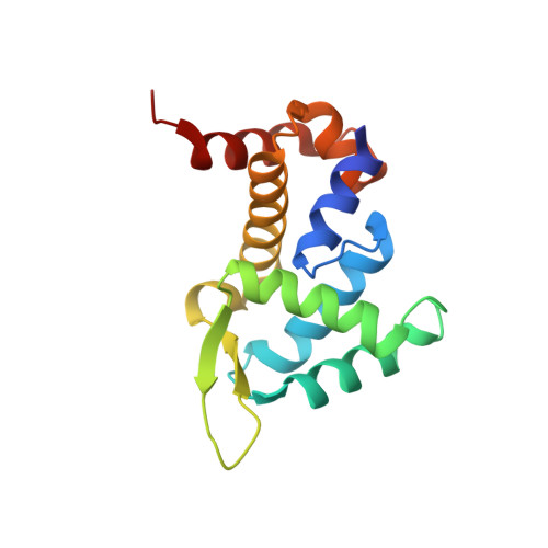
Roquin Binding to Target Mrnas Involves a Winged Helix-Turn- Helix Motif.

Schuetz, A.Murakawa, Y.Rosenbaum, E.Landthaler, M.Heinemann, U.

(2014) Nat Commun 5: 5701

  • DOI: https://doi.org/10.1038/ncomms6701
  • Primary Citation of Related Structures:  
    4ULW

  • PubMed Abstract: 

    Roquin proteins mediate mRNA deadenylation by recognizing a conserved class of stem-loop RNA degradation motifs via their Roquin domain. Here we present the crystal structure of a Roquin domain, revealing a mostly helical protein fold bearing a winged helix-turn-helix motif. By combining structural, biochemical and mutation analyses, we gain insight into the mode of RNA binding. We show that the winged helix-turn-helix motif is involved in the binding of constitutive decay elements-containing stem-loop mRNAs. Moreover, we provide biochemical evidence that Roquin proteins are additionally able to bind to duplex RNA and have the potential to be functional in different oligomeric states.


  • Organizational Affiliation

    Helmholtz Protein Sample Production Facility, Max Delbrück Center for Molecular Medicine, 13092 Berlin, Germany.


Macromolecules
Find similar proteins by:  (by identity cutoff)  |  3D Structure
Entity ID: 1
MoleculeChains Sequence LengthOrganismDetailsImage
ROQUIN-1
A, B
152Homo sapiensMutation(s): 0 
UniProt & NIH Common Fund Data Resources
Find proteins for Q5TC82 (Homo sapiens)
Explore Q5TC82 
Go to UniProtKB:  Q5TC82
PHAROS:  Q5TC82
GTEx:  ENSG00000135870 
Entity Groups  
Sequence Clusters30% Identity50% Identity70% Identity90% Identity95% Identity100% Identity
UniProt GroupQ5TC82
Sequence Annotations
Expand
  • Reference Sequence
Experimental Data & Validation

Experimental Data

  • Method: X-RAY DIFFRACTION
  • Resolution: 1.91 Å
  • R-Value Free: 0.241 
  • R-Value Work: 0.200 
  • R-Value Observed: 0.202 
  • Space Group: C 1 2 1
Unit Cell:
Length ( Å )Angle ( ˚ )
a = 169.66α = 90
b = 29.785β = 100.94
c = 60.197γ = 90
Software Package:
Software NamePurpose
REFMACrefinement

Structure Validation

View Full Validation Report


Entry History 

Deposition Data

Revision History  (Full details and data files)

  • Version 1.0: 2015-01-14
    Type: Initial release