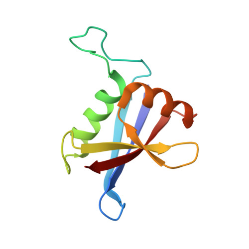
4UF8

Electron cryo-microscopy structure of PB1-p62 filaments


Experimental Data Snapshot

  • Method: ELECTRON MICROSCOPY
  • Resolution: 10.9 Å
  • Aggregation State: FILAMENT 
  • Reconstruction Method: HELICAL 

Starting Model: experimental
View more details

wwPDB Validation   3D Report Full Report


This is version 1.3 of the entry. See complete history


Literature

The Selective Autophagy Receptor P62 Forms a Flexible Filamentous Helical Scaffold.

Ciuffa, R.Lamark, T.Tarafder, A.K.Guesdon, A.Rybina, S.Hagen, W.J.H.Johansen, T.Sachse, C.

(2015) Cell Rep 11: 748

  • DOI: https://doi.org/10.1016/j.celrep.2015.03.062
  • Primary Citation of Related Structures:  
    4UF8, 4UF9

  • PubMed Abstract: 

    The scaffold protein p62/SQSTM1 is involved in protein turnover and signaling and is commonly found in dense protein bodies in eukaryotic cells. In autophagy, p62 acts as a selective autophagy receptor that recognizes and shuttles ubiquitinated proteins to the autophagosome for degradation. The structural organization of p62 in cellular bodies and the interplay of these assemblies with ubiquitin and the autophagic marker LC3 remain to be elucidated. Here, we present a cryo-EM structural analysis of p62. Together with structures of assemblies from the PB1 domain, we show that p62 is organized in flexible polymers with the PB1 domain constituting a helical scaffold. Filamentous p62 is capable of binding LC3 and addition of long ubiquitin chains induces disassembly and shortening of filaments. These studies explain how p62 assemblies provide a large molecular scaffold for the nascent autophagosome and reveal how they can bind ubiquitinated cargo.


  • Organizational Affiliation

    European Molecular Biology Laboratory, Structural and Computation Biology Unit, Meyerhofstrasse 1, 69117 Heidelberg, Germany.


Macromolecules
Find similar proteins by:  (by identity cutoff)  |  3D Structure
Entity ID: 1
MoleculeChains Sequence LengthOrganismDetailsImage
SEQUESTOSOME-1A,
B,
C,
D [auth I]
100Homo sapiensMutation(s): 0 
UniProt & NIH Common Fund Data Resources
Find proteins for Q13501 (Homo sapiens)
Explore Q13501 
Go to UniProtKB:  Q13501
PHAROS:  Q13501
GTEx:  ENSG00000161011 
Entity Groups  
Sequence Clusters30% Identity50% Identity70% Identity90% Identity95% Identity100% Identity
UniProt GroupQ13501
Sequence Annotations
Expand
  • Reference Sequence
Experimental Data & Validation

Experimental Data

  • Method: ELECTRON MICROSCOPY
  • Resolution: 10.9 Å
  • Aggregation State: FILAMENT 
  • Reconstruction Method: HELICAL 
EM Software:
TaskSoftware PackageVersion
RECONSTRUCTIONSPRING

Structure Validation

View Full Validation Report


Entry History 

Deposition Data

Revision History  (Full details and data files)

  • Version 1.0: 2015-05-13
    Type: Initial release
  • Version 1.1: 2015-05-27
    Changes: Database references
  • Version 1.2: 2017-08-23
    Changes: Data collection
  • Version 1.3: 2024-05-08
    Changes: Data collection, Database references, Derived calculations, Refinement description