4UBV | pdb_00004ubv

Structure of the 3-ketoacyl-CoA thiolase FadA5 from M. tuberculosis with an partially acetylated cysteine in complex with acetyl-CoA and CoA


Experimental Data Snapshot

  • Method: X-RAY DIFFRACTION
  • Resolution: 1.95 Å
  • R-Value Free: 
    0.231 (Depositor), 0.197 (DCC) 
  • R-Value Work: 
    0.193 (Depositor) 
  • R-Value Observed: 
    0.195 (Depositor) 

Starting Model: experimental
View more details

wwPDB Validation 3D Report Full Report

Validation slider image for 4UBV

Ligand Structure Quality Assessment 


This is version 2.2 of the entry. See complete history

Literature

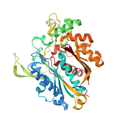
FadA5 a Thiolase from Mycobacterium tuberculosis: A Steroid-Binding Pocket Reveals the Potential for Drug Development against Tuberculosis.

Schaefer, C.M.Lu, R.Nesbitt, N.M.Schiebel, J.Sampson, N.S.Kisker, C.

(2015) Structure 23: 21-33

  • DOI: https://doi.org/10.1016/j.str.2014.10.010
  • Primary Citation Related Structures: 
    4UBT, 4UBU, 4UBV, 4UBW

  • PubMed Abstract: 

    With the exception of HIV, tuberculosis (TB) is the leading cause of mortality among infectious diseases. The urgent need to develop new antitubercular drugs is apparent due to the increasing number of drug-resistant Mycobacterium tuberculosis (Mtb) strains. Proteins involved in cholesterol import and metabolism have recently been discovered as potent targets against TB. FadA5, a thiolase from Mtb, is catalyzing the last step of the β-oxidation reaction of the cholesterol side-chain degradation under release of critical metabolites and was shown to be of importance during the chronic stage of TB infections. To gain structural and mechanistic insight on FadA5, we characterized the enzyme in different stages of the cleavage reaction and with a steroid bound to the binding pocket. Structural comparisons to human thiolases revealed that it should be possible to target FadA5 specifically, and the steroid-bound structure provides a solid basis for the development of inhibitors against FadA5.


  • Organizational Affiliation
    • Rudolf Virchow Center for Experimental Biomedicine, Institute for Structural Biology, University of Würzburg, Josef-Schneider-Strasse 2, 97080 Würzburg, Germany.

Macromolecule Content 

  • Total Structure Weight: 89.05 kDa 
  • Atom Count: 6,504 
  • Modeled Residue Count: 785 
  • Deposited Residue Count: 800 
  • Unique protein chains: 1

Macromolecules

Find similar proteins by:|  3D Structure
Entity ID: 1
MoleculeChains  Sequence LengthOrganismDetailsImage
Acetyl-CoA acetyltransferase FadA5
A, B
399Mycobacterium tuberculosis H37RvMutation(s): 0 
Gene Names: fadA5Rv3546P425_03688RVBD_3546
EC: 2.3.1.16
UniProt
Find proteins for I6XHI4 (Mycobacterium tuberculosis (strain ATCC 25618 / H37Rv))
Explore I6XHI4 
Go to UniProtKB:  I6XHI4
Entity Groups
Sequence Clusters30% Identity50% Identity70% Identity90% Identity95% Identity100% Identity
UniProt GroupI6XHI4
Sequence Annotations
Expand
Reference Sequence

Small Molecules

Ligands 5 Unique
IDChains Name / Formula / InChI Key2D Diagram3D Interactions
ACO

Query on ACO


Download:Ideal Coordinates CCD File
C [auth A]ACETYL COENZYME *A
C23 H38 N7 O17 P3 S
ZSLZBFCDCINBPY-ZSJPKINUSA-N
COA

Query on COA


Download:Ideal Coordinates CCD File
D [auth A],
M [auth B]
COENZYME A
C21 H36 N7 O16 P3 S
RGJOEKWQDUBAIZ-IBOSZNHHSA-N
SO4

Query on SO4


Download:Ideal Coordinates CCD File
E [auth A],
F [auth A],
N [auth B]
SULFATE ION
O4 S
QAOWNCQODCNURD-UHFFFAOYSA-L
GOL

Query on GOL


Download:Ideal Coordinates CCD File
AA [auth B]
I [auth A]
J [auth A]
K [auth A]
L [auth A]
AA [auth B],
I [auth A],
J [auth A],
K [auth A],
L [auth A],
T [auth B],
U [auth B],
V [auth B],
W [auth B],
X [auth B],
Y [auth B],
Z [auth B]
GLYCEROL
C3 H8 O3
PEDCQBHIVMGVHV-UHFFFAOYSA-N
DIO

Query on DIO


Download:Ideal Coordinates CCD File
G [auth A]
H [auth A]
O [auth B]
P [auth B]
Q [auth B]
G [auth A],
H [auth A],
O [auth B],
P [auth B],
Q [auth B],
R [auth B],
S [auth B]
1,4-DIETHYLENE DIOXIDE
C4 H8 O2
RYHBNJHYFVUHQT-UHFFFAOYSA-N
Modified Residues  1 Unique
IDChains TypeFormula2D DiagramParent
SCY
Query on SCY
A, B
L-PEPTIDE LINKINGC5 H9 N O3 SCYS

Experimental Data & Validation

Experimental Data

  • Method: X-RAY DIFFRACTION
  • Resolution: 1.95 Å
  • R-Value Free:  0.231 (Depositor), 0.197 (DCC) 
  • R-Value Work:  0.193 (Depositor) 
  • R-Value Observed: 0.195 (Depositor) 
Space Group: P 43 21 2
Unit Cell:
Length ( Å )Angle ( ˚ )
a = 124.68α = 90
b = 124.68β = 90
c = 124.71γ = 90
Software Package:
Software NamePurpose
REFMACrefinement
SCALAdata scaling
iMOSFLMdata reduction
PHASERphasing

Structure Validation

View Full Validation Report


Ligand Structure Quality Assessment 


Entry History 

& Funding Information

Deposition Data


Funding OrganizationLocationGrant Number
German Research FoundationGermanySFB630
German Research FoundationGermanyFZ82
National Institutes of HealthUnited StatesAI092455
National Institutes of HealthUnited StatesAI085349
National Institutes of HealthUnited StatesAI065251
National Institutes of HealthUnited StatesHL53306
National Institutes of HealthUnited StatesRR021008
NSFUnited StatesBIO1039771

Revision History  (Full details and data files)

  • Version 1.0: 2014-12-17
    Type: Initial release
  • Version 1.1: 2015-01-14
    Changes: Database references
  • Version 2.0: 2021-09-08
    Changes: Advisory, Atomic model, Author supporting evidence, Data collection, Database references, Derived calculations, Polymer sequence, Source and taxonomy, Structure summary
  • Version 2.1: 2023-12-20
    Changes: Data collection, Refinement description
  • Version 2.2: 2024-11-13
    Changes: Structure summary