4UBF | pdb_00004ubf

HsMCAK motor domain complex


Experimental Data Snapshot

  • Method: X-RAY DIFFRACTION
  • Resolution: 3.00 Å
  • R-Value Free: 
    0.286 (Depositor), 0.308 (DCC) 
  • R-Value Work: 
    0.264 (Depositor) 
  • R-Value Observed: 
    0.265 (Depositor) 

Starting Model: experimental
View more details

wwPDB Validation 3D Report Full Report

Validation slider image for 4UBF

Ligand Structure Quality Assessment 


This is version 1.3 of the entry. See complete history

Literature

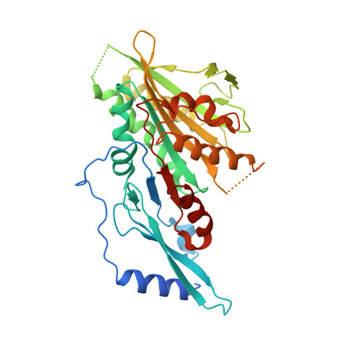
The C-terminal region of the motor protein MCAK controls its structure and activity through a conformational switch.

Talapatra, S.K.Harker, B.Welburn, J.P.

(2015) Elife 4

  • DOI: https://doi.org/10.7554/eLife.06421
  • Primary Citation Related Structures: 
    4UBF

  • PubMed Abstract: 

    The precise regulation of microtubule dynamics is essential during cell division. The kinesin-13 motor protein MCAK is a potent microtubule depolymerase. The divergent non-motor regions flanking the ATPase domain are critical in regulating its targeting and activity. However, the molecular basis for the function of the non-motor regions within the context of full-length MCAK is unknown. Here, we determine the structure of MCAK motor domain bound to its regulatory C-terminus. Our analysis reveals that the MCAK C-terminus binds to two motor domains in solution and is displaced allosterically upon microtubule binding, which allows its robust accumulation at microtubule ends. These results demonstrate that MCAK undergoes long-range conformational changes involving its C-terminus during the soluble to microtubule-bound transition and that the C-terminus-motor interaction represents a structural intermediate in the MCAK catalytic cycle. Together, our work reveals intrinsic molecular mechanisms underlying the regulation of kinesin-13 activity.


  • Organizational Affiliation
    • Wellcome Trust Centre for Cell Biology, School of Biological Sciences, University of Edinburgh, Edinburgh, United Kingdom.

Macromolecule Content 

  • Total Structure Weight: 176.44 kDa 
  • Atom Count: 9,707 
  • Modeled Residue Count: 1,279 
  • Deposited Residue Count: 1,560 
  • Unique protein chains: 2

Macromolecules

Find similar proteins by:|  3D Structure
Entity ID: 1
MoleculeChains  Sequence LengthOrganismDetailsImage
Kinesin-like protein KIF2C
A, B, C, D
387Homo sapiensMutation(s): 0 
Gene Names: KIF2CKNSL6
UniProt & NIH Common Fund Data Resources
Find proteins for Q99661 (Homo sapiens)
Explore Q99661 
Go to UniProtKB:  Q99661
PHAROS:  Q99661
GTEx:  ENSG00000142945 
Entity Groups
Sequence Clusters30% Identity50% Identity70% Identity90% Identity95% Identity100% Identity
UniProt GroupQ99661
Sequence Annotations
Expand
Reference Sequence
Find similar proteins by:  Sequence   |   3D Structure  
Entity ID: 2
MoleculeChains  Sequence LengthOrganismDetailsImage
Kinesin-like protein KIF2CE [auth P]12Homo sapiensMutation(s): 0 
Gene Names: KIF2CKNSL6
UniProt & NIH Common Fund Data Resources
Find proteins for Q99661 (Homo sapiens)
Explore Q99661 
Go to UniProtKB:  Q99661
PHAROS:  Q99661
GTEx:  ENSG00000142945 
Entity Groups
Sequence Clusters30% Identity50% Identity70% Identity90% Identity95% Identity100% Identity
UniProt GroupQ99661
Sequence Annotations
Expand
Reference Sequence

Small Molecules

Ligands 2 Unique
IDChains Name / Formula / InChI Key2D Diagram3D Interactions
ADP

Query on ADP


Download:Ideal Coordinates CCD File
G [auth A],
I [auth B],
M [auth C],
P [auth D]
ADENOSINE-5'-DIPHOSPHATE
C10 H15 N5 O10 P2
XTWYTFMLZFPYCI-KQYNXXCUSA-N
MG

Query on MG


Download:Ideal Coordinates CCD File
F [auth A]
H [auth A]
J [auth B]
K [auth B]
L [auth C]
F [auth A],
H [auth A],
J [auth B],
K [auth B],
L [auth C],
N [auth C],
O [auth D]
MAGNESIUM ION
Mg
JLVVSXFLKOJNIY-UHFFFAOYSA-N

Experimental Data & Validation

Experimental Data

  • Method: X-RAY DIFFRACTION
  • Resolution: 3.00 Å
  • R-Value Free:  0.286 (Depositor), 0.308 (DCC) 
  • R-Value Work:  0.264 (Depositor) 
  • R-Value Observed: 0.265 (Depositor) 
Space Group: P 1 21 1
Unit Cell:
Length ( Å )Angle ( ˚ )
a = 46.306α = 90
b = 245.644β = 95.84
c = 79.399γ = 90
Software Package:
Software NamePurpose
PHENIXrefinement

Structure Validation

View Full Validation Report


Ligand Structure Quality Assessment 


Entry History 

& Funding Information

Deposition Data


Funding OrganizationLocationGrant Number
Cancer Research UKUnited Kingdom543WJW_R42094

Revision History  (Full details and data files)

  • Version 1.0: 2015-05-06
    Type: Initial release
  • Version 1.1: 2017-08-30
    Changes: Author supporting evidence
  • Version 1.2: 2018-06-06
    Changes: Data collection, Database references
  • Version 1.3: 2023-12-20
    Changes: Data collection, Database references, Derived calculations, Refinement description