4UB2

DNA polymerase beta product complex with a templating cytosine and 8-oxodGMP, 120 s


Experimental Data Snapshot

  • Method: X-RAY DIFFRACTION
  • Resolution: 2.51 Å
  • R-Value Free: 0.292 
  • R-Value Work: 0.226 
  • R-Value Observed: 0.229 

wwPDB Validation   3D Report Full Report


This is version 1.4 of the entry. See complete history


Literature

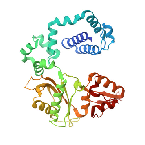
Uncovering the polymerase-induced cytotoxicity of an oxidized nucleotide.

Freudenthal, B.D.Beard, W.A.Perera, L.Shock, D.D.Kim, T.Schlick, T.Wilson, S.H.

(2015) Nature 517: 635-639

  • DOI: https://doi.org/10.1038/nature13886
  • Primary Citation of Related Structures:  
    4UAW, 4UAY, 4UAZ, 4UB1, 4UB2, 4UB3, 4UB4, 4UB5, 4UBB, 4UBC

  • PubMed Abstract: 

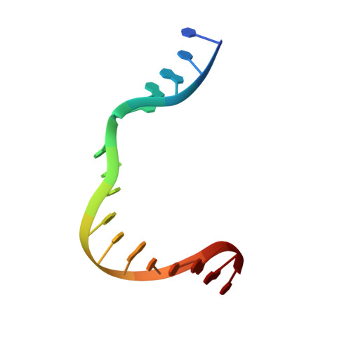
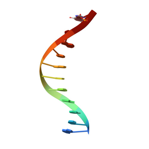
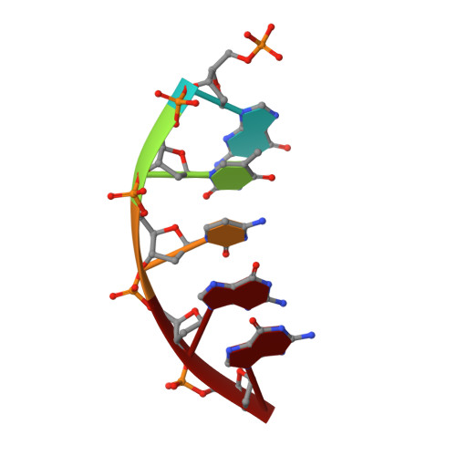
    Oxidative stress promotes genomic instability and human diseases. A common oxidized nucleoside is 8-oxo-7,8-dihydro-2'-deoxyguanosine, which is found both in DNA (8-oxo-G) and as a free nucleotide (8-oxo-dGTP). Nucleotide pools are especially vulnerable to oxidative damage. Therefore cells encode an enzyme (MutT/MTH1) that removes free oxidized nucleotides. This cleansing function is required for cancer cell survival and to modulate Escherichia coli antibiotic sensitivity in a DNA polymerase (pol)-dependent manner. How polymerases discriminate between damaged and non-damaged nucleotides is not well understood. This analysis is essential given the role of oxidized nucleotides in mutagenesis, cancer therapeutics, and bacterial antibiotics. Even with cellular sanitizing activities, nucleotide pools contain enough 8-oxo-dGTP to promote mutagenesis. This arises from the dual coding potential where 8-oxo-dGTP(anti) base pairs with cytosine and 8-oxo-dGTP(syn) uses its Hoogsteen edge to base pair with adenine. Here we use time-lapse crystallography to follow 8-oxo-dGTP insertion opposite adenine or cytosine with human pol β, to reveal that insertion is accommodated in either the syn- or anti-conformation, respectively. For 8-oxo-dGTP(anti) insertion, a novel divalent metal relieves repulsive interactions between the adducted guanine base and the triphosphate of the oxidized nucleotide. With either templating base, hydrogen-bonding interactions between the bases are lost as the enzyme reopens after catalysis, leading to a cytotoxic nicked DNA repair intermediate. Combining structural snapshots with kinetic and computational analysis reveals how 8-oxo-dGTP uses charge modulation during insertion that can lead to a blocked DNA repair intermediate.


  • Organizational Affiliation

    Laboratory of Structural Biology, National Institute of Environmental Health Sciences, National Institutes of Health, PO Box 12233, Research Triangle Park, North Carolina 27709-2233, USA.


Macromolecules

Find similar proteins by:  (by identity cutoff)  |  3D Structure
Entity ID: 1
MoleculeChains Sequence LengthOrganismDetailsImage
DNA polymerase beta335Homo sapiensMutation(s): 0 
Gene Names: POLB
EC: 2.7.7.7 (PDB Primary Data), 4.2.99 (PDB Primary Data)
UniProt & NIH Common Fund Data Resources
Find proteins for P06746 (Homo sapiens)
Explore P06746 
Go to UniProtKB:  P06746
PHAROS:  P06746
GTEx:  ENSG00000070501 
Entity Groups  
Sequence Clusters30% Identity50% Identity70% Identity90% Identity95% Identity100% Identity
UniProt GroupP06746
Sequence Annotations
Expand
  • Reference Sequence

Find similar nucleic acids by:  Sequence   |   3D Structure  

Entity ID: 2
MoleculeChains LengthOrganismImage
5'-D(*CP*CP*GP*AP*CP*CP*GP*CP*GP*CP*AP*TP*CP*AP*GP*C)-3'B [auth T]16synthetic construct
Sequence Annotations
Expand
  • Reference Sequence

Find similar nucleic acids by:  Sequence   |   3D Structure  

Entity ID: 3
MoleculeChains LengthOrganismImage
5'-D(*GP*CP*TP*GP*AP*TP*GP*CP*GP*CP*(8OG))-3'C [auth P]11synthetic construct
Sequence Annotations
Expand
  • Reference Sequence

Find similar nucleic acids by:  Sequence   |   3D Structure  

Entity ID: 4
MoleculeChains LengthOrganismImage
5'-D(P*GP*TP*CP*GP*G)-3'5synthetic construct
Sequence Annotations
Expand
  • Reference Sequence
Experimental Data & Validation

Experimental Data

  • Method: X-RAY DIFFRACTION
  • Resolution: 2.51 Å
  • R-Value Free: 0.292 
  • R-Value Work: 0.226 
  • R-Value Observed: 0.229 
  • Space Group: P 1 21 1
Unit Cell:
Length ( Å )Angle ( ˚ )
a = 55.099α = 90
b = 79.589β = 109.69
c = 55.668γ = 90
Software Package:
Software NamePurpose
PHENIXrefinement

Structure Validation

View Full Validation Report


Entry History & Funding Information

Deposition Data


Funding OrganizationLocationGrant Number
National Institutes of Health/National Institute of Environmental Health Sciences (NIH/NIEHS)United StatesES050158
National Institutes of Health/National Institute of Environmental Health Sciences (NIH/NIEHS)United StatesES050161
National Institutes of Health/National Institute of Environmental Health Sciences (NIH/NIEHS)United StatesES043010

Revision History  (Full details and data files)

  • Version 1.0: 2014-11-12
    Type: Initial release
  • Version 1.1: 2014-12-03
    Changes: Database references
  • Version 1.2: 2015-02-04
    Changes: Database references
  • Version 1.3: 2019-11-27
    Changes: Author supporting evidence, Database references, Derived calculations, Other, Source and taxonomy
  • Version 1.4: 2023-09-27
    Changes: Data collection, Database references, Refinement description