4UAM | pdb_00004uam

1.8 Angstrom crystal structure of IMP-1 metallo-beta-lactamase with a mixed iron-zinc center in the active site


Experimental Data Snapshot

  • Method: X-RAY DIFFRACTION
  • Resolution: 1.80 Å
  • R-Value Free: 
    0.182 (Depositor), 0.184 (DCC) 
  • R-Value Work: 
    0.162 (Depositor), 0.164 (DCC) 
  • R-Value Observed: 
    0.163 (Depositor) 

Starting Model: experimental
View more details

wwPDB Validation 3D Report Full Report

Validation slider image for 4UAM

This is version 1.7 of the entry. See complete history

Literature

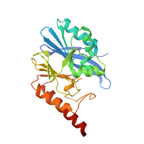
Iron(III) Located in the Dinuclear Metallo-beta-Lactamase IMP-1 by Pseudocontact Shifts.

Carruthers, T.J.Carr, P.D.Loh, C.T.Jackson, C.J.Otting, G.

(2014) Angew Chem Int Ed Engl 53: 14269-14272

  • DOI: https://doi.org/10.1002/anie.201408693
  • Primary Citation Related Structures: 
    4UAM

  • PubMed Abstract: 

    Heterodinuclear metalloenzymes are an important class of metalloproteins, but determining the location of the different metal ions can be difficult. Herein we present a new NMR spectroscopy method that uses pseudocontact shifts (PCS) to achieve this without assumptions about the coordinating ligands. The approach is illustrated with the dinuclear [FeZn] complex of IMP-1, which is a prototypical metallo-β-lactamase (MβL) that confers resistance to β-lactam antibiotics. Results from single-crystal X-ray diffraction were compromised by degradation during crystallization. With [GaZn]-IMP-1 as diamagnetic reference, the PCSs unambiguously identified the iron binding site in fresh samples of [FeZn]-IMP-1, even though the two metal centers are less than 3.8 Å apart and the iron is high-spin Fe(3+), which produces only small PCSs. [FeZn]-MβLs may be important drug targets, as [FeZn]-IMP-1 is enzymatically active and readily produced in the presence of small amounts of Fe(3+).


  • Organizational Affiliation
    • Research School of Chemistry, Australian National University, Canberra, ACT 0200 (Australia).

Macromolecule Content 

  • Total Structure Weight: 101.83 kDa 
  • Atom Count: 7,961 
  • Modeled Residue Count: 884 
  • Deposited Residue Count: 916 
  • Unique protein chains: 1

Macromolecules

Find similar proteins by:|  3D Structure
Entity ID: 1
MoleculeChains  Sequence LengthOrganismDetailsImage
IMP-1 metallo-beta-lactamase
A, B, C, D
228Pseudomonas aeruginosa NCGM2.S1Mutation(s): 1 
Gene Names: bla IMPNCGM2_1757

Small Molecules

Ligands 3 Unique
IDChains Name / Formula / InChI Key2D Diagram3D Interactions
FLC

Query on FLC


Download:Ideal Coordinates CCD File
G [auth A],
J [auth B],
M [auth C],
P [auth D]
CITRATE ANION
C6 H5 O7
KRKNYBCHXYNGOX-UHFFFAOYSA-K
ZN

Query on ZN


Download:Ideal Coordinates CCD File
F [auth A],
I [auth B],
L [auth C],
O [auth D]
ZINC ION
Zn
PTFCDOFLOPIGGS-UHFFFAOYSA-N
FE

Query on FE


Download:Ideal Coordinates CCD File
E [auth A],
H [auth B],
K [auth C],
N [auth D]
FE (III) ION
Fe
VTLYFUHAOXGGBS-UHFFFAOYSA-N
Modified Residues  1 Unique
IDChains TypeFormula2D DiagramParent
CSD
Query on CSD
A, B, C, D
L-PEPTIDE LINKINGC3 H7 N O4 SCYS

Experimental Data & Validation

Experimental Data

  • Method: X-RAY DIFFRACTION
  • Resolution: 1.80 Å
  • R-Value Free:  0.182 (Depositor), 0.184 (DCC) 
  • R-Value Work:  0.162 (Depositor), 0.164 (DCC) 
  • R-Value Observed: 0.163 (Depositor) 
Space Group: P 1
Unit Cell:
Length ( Å )Angle ( ˚ )
a = 49.99α = 83.45
b = 75.91β = 75.3
c = 82.36γ = 74.01
Software Package:
Software NamePurpose
PDB_EXTRACTdata extraction
PHENIXrefinement
Cootmodel building
PHASERphasing
XSCALEdata scaling
XDSdata reduction
Blu-Icedata collection

Structure Validation

View Full Validation Report


Entry History 

Deposition Data

Revision History  (Full details and data files)

  • Version 1.0: 2014-09-17
    Type: Initial release
  • Version 1.1: 2014-11-05
    Changes: Database references
  • Version 1.2: 2014-11-12
    Changes: Structure summary
  • Version 1.3: 2014-12-24
    Changes: Database references
  • Version 1.4: 2017-08-23
    Changes: Data collection, Database references, Derived calculations, Other, Refinement description, Source and taxonomy, Structure summary
  • Version 1.5: 2017-09-06
    Changes: Author supporting evidence
  • Version 1.6: 2023-09-27
    Changes: Data collection, Database references, Derived calculations, Refinement description
  • Version 1.7: 2024-10-16
    Changes: Structure summary