4U8W | pdb_00004u8w

HIV-1 wild Type protease with GRL-050-10A (a Gem-difluoro-bis-Tetrahydrofuran as P2-Ligand)


Experimental Data Snapshot

  • Method: X-RAY DIFFRACTION
  • Resolution: 1.30 Å
  • R-Value Free: 
    0.188 (Depositor), 0.183 (DCC) 
  • R-Value Work: 
    0.154 (Depositor), 0.154 (DCC) 
  • R-Value Observed: 
    0.156 (Depositor) 

Starting Model: experimental
View more details

wwPDB Validation 3D Report Full Report

Validation slider image for 4U8W

Ligand Structure Quality Assessment 


This is version 1.4 of the entry. See complete history

Literature

Design of gem-Difluoro-bis-Tetrahydrofuran as P2 Ligand for HIV-1 Protease Inhibitors to Improve Brain Penetration: Synthesis, X-ray Studies, and Biological Evaluation.

Ghosh, A.K.Yashchuk, S.Mizuno, A.Chakraborty, N.Agniswamy, J.Wang, Y.F.Aoki, M.Gomez, P.M.Amano, M.Weber, I.T.Mitsuya, H.

(2015) ChemMedChem 10: 107-115

  • DOI: https://doi.org/10.1002/cmdc.201402358
  • Primary Citation Related Structures: 
    4U8W

  • PubMed Abstract: 

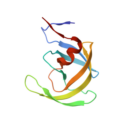
    The structure-based design, synthesis, biological evaluation, and X-ray structural studies of fluorine-containing HIV-1 protease inhibitors are described. The synthesis of both enantiomers of the gem-difluoro-bis-THF ligands was carried out in a stereoselective manner using a Reformatskii-Claisen reaction as the key step. Optically active ligands were converted into protease inhibitors. Two of these inhibitors, (3R,3aS,6aS)-4,4-difluorohexahydrofuro[2,3-b]furan-3-yl(2S,3R)-3-hydroxy-4-((N-isobutyl-4-methoxyphenyl)sulfonamido)-1-phenylbutan-2-yl) carbamate (3) and (3R,3aS,6aS)-4,4-difluorohexahydrofuro[2,3-b]furan-3-yl(2S,3R)-3-hydroxy-4-((N-isobutyl-4-aminophenyl)sulfonamido)phenylbutan-2-yl) carbamate (4), exhibited HIV-1 protease inhibitory Ki values in the picomolar range. Both 3 and 4 showed very potent antiviral activity, with respective EC50 values of 0.8 and 3.1 nM against the laboratory strain HIV-1LAI . The two inhibitors exhibited better lipophilicity profiles than darunavir, and also showed much improved blood-brain barrier permeability in an in vitro model. A high-resolution X-ray structure of inhibitor 4 in complex with HIV-1 protease was determined, revealing that the fluorinated ligand makes extensive interactions with the S2 subsite of HIV-1 protease, including hydrogen bonding interactions with the protease backbone atoms. Moreover, both fluorine atoms on the bis-THF ligand formed strong interactions with the flap Gly 48 carbonyl oxygen atom.


  • Organizational Affiliation
    • Departments of Chemistry and Medicinal Chemistry, Purdue University, West Lafayette, IN 47907 (USA). akghosh@purdue.edu.

Macromolecule Content 

  • Total Structure Weight: 22.31 kDa 
  • Atom Count: 1,971 
  • Modeled Residue Count: 198 
  • Deposited Residue Count: 198 
  • Unique protein chains: 1

Macromolecules

Find similar proteins by:|  3D Structure
Entity ID: 1
MoleculeChains  Sequence LengthOrganismDetailsImage
HIV-1 Protease
A, B
99Human immunodeficiency virus type 1 BH10Mutation(s): 5 
Gene Names: gag-pol
EC: 3.4.23.16
UniProt
Find proteins for P03366 (Human immunodeficiency virus type 1 group M subtype B (isolate BH10))
Explore P03366 
Go to UniProtKB:  P03366
Entity Groups
Sequence Clusters30% Identity50% Identity70% Identity90% Identity95% Identity100% Identity
UniProt GroupP03366
Sequence Annotations
Expand
Reference Sequence

Small Molecules

Ligands 4 Unique
IDChains Name / Formula / InChI Key2D Diagram3D Interactions
G10

Query on G10


Download:Ideal Coordinates CCD File
C [auth A](3R,3aS,6aS)-4,4-difluorohexahydrofuro[2,3-b]furan-3-yl [(2S,3R)-4-{[(4-aminophenyl)sulfonyl](2-methylpropyl)amino}-3-hydroxy-1-phenylbutan-2-yl]carbamate
C27 H35 F2 N3 O7 S
UQYAKJSDOQPRAX-YCXOGWGTSA-N
ACT

Query on ACT


Download:Ideal Coordinates CCD File
H [auth B],
I [auth B]
ACETATE ION
C2 H3 O2
QTBSBXVTEAMEQO-UHFFFAOYSA-M
CL

Query on CL


Download:Ideal Coordinates CCD File
E [auth A],
F [auth B],
G [auth B]
CHLORIDE ION
Cl
VEXZGXHMUGYJMC-UHFFFAOYSA-M
NA

Query on NA


Download:Ideal Coordinates CCD File
D [auth A]SODIUM ION
Na
FKNQFGJONOIPTF-UHFFFAOYSA-N

Experimental Data & Validation

Experimental Data

  • Method: X-RAY DIFFRACTION
  • Resolution: 1.30 Å
  • R-Value Free:  0.188 (Depositor), 0.183 (DCC) 
  • R-Value Work:  0.154 (Depositor), 0.154 (DCC) 
  • R-Value Observed: 0.156 (Depositor) 
Space Group: P 21 21 2
Unit Cell:
Length ( Å )Angle ( ˚ )
a = 58.539α = 90
b = 85.989β = 90
c = 46.21γ = 90
Software Package:
Software NamePurpose
SHELXL-97refinement
HKL-2000data collection
HKL-2000data scaling
PHASERphasing
HKL-2000data reduction

Structure Validation

View Full Validation Report


Ligand Structure Quality Assessment 


Entry History 

& Funding Information

Deposition Data


Funding OrganizationLocationGrant Number
National Institutes of Health/National Institute of General Medical Sciences (NIH/NIGMS)United StatesGM62920

Revision History  (Full details and data files)

  • Version 1.0: 2014-11-05
    Type: Initial release
  • Version 1.1: 2014-12-31
    Changes: Database references
  • Version 1.2: 2017-09-27
    Changes: Author supporting evidence, Derived calculations, Other, Refinement description, Source and taxonomy
  • Version 1.3: 2019-12-25
    Changes: Author supporting evidence
  • Version 1.4: 2023-09-27
    Changes: Data collection, Database references, Derived calculations, Refinement description