4U8T

Crystal structure of YTH domain of Zygosaccharomyces rouxii MRB1 protein in complex with N6-Methyladenosine RNA


Experimental Data Snapshot

  • Method: X-RAY DIFFRACTION
  • Resolution: 2.70 Å
  • R-Value Free: 0.245 
  • R-Value Work: 0.199 
  • R-Value Observed: 0.201 

wwPDB Validation   3D Report Full Report


This is version 2.0 of the entry. See complete history


Literature

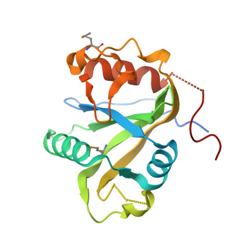
Molecular basis for the recognition of methylated adenines in RNA by the eukaryotic YTH domain.

Luo, S.Tong, L.

(2014) Proc Natl Acad Sci U S A 111: 13834-13839

  • DOI: https://doi.org/10.1073/pnas.1412742111
  • Primary Citation of Related Structures:  
    4U8T

  • PubMed Abstract: 

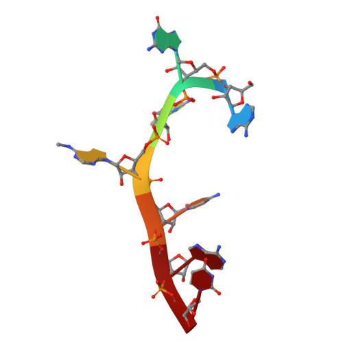
    Methylation of the N6 position of selected internal adenines (m(6)A) in mRNAs and noncoding RNAs is widespread in eukaryotes, and the YTH domain in a collection of proteins recognizes this modification. We report the crystal structure of the splicing factor YT521-B homology (YTH) domain of Zygosaccharomyces rouxii MRB1 in complex with a heptaribonucleotide with an m(6)A residue in the center. The m(6)A modification is recognized by an aromatic cage, being sandwiched between a Trp and Tyr residue and with the methyl group pointed toward another Trp residue. Mutations of YTH domain residues in the RNA binding site can abolish the formation of the complex, confirming the structural observations. These residues are conserved in the human YTH proteins that also bind m(6)A RNA, suggesting a conserved mode of recognition. Overall, our structural and biochemical studies have defined the molecular basis for how the YTH domain functions as a reader of methylated adenines.


  • Organizational Affiliation

    Department of Biological Sciences, Columbia University, New York, NY 10027.


Macromolecules

Find similar proteins by:  (by identity cutoff)  |  3D Structure
Entity ID: 1
MoleculeChains Sequence LengthOrganismDetailsImage
ZYRO0G01672p
A, B, C, D, E
A, B, C, D, E, F
177Zygosaccharomyces rouxii CBS 732Mutation(s): 0 
Gene Names: ZYRO0G01672g
UniProt
Find proteins for C5E1V0 (Zygosaccharomyces rouxii (strain ATCC 2623 / CBS 732 / NBRC 1130 / NCYC 568 / NRRL Y-229))
Explore C5E1V0 
Go to UniProtKB:  C5E1V0
Entity Groups  
Sequence Clusters30% Identity50% Identity70% Identity90% Identity95% Identity100% Identity
UniProt GroupC5E1V0
Sequence Annotations
Expand
  • Reference Sequence

Find similar nucleic acids by:  Sequence   |   3D Structure  

Entity ID: 2
MoleculeChains LengthOrganismImage
RNA (5'-R(*AP*GP*GP*(6MZ)P*CP*AP*U)-3')7Saccharomyces cerevisiae
Sequence Annotations
Expand
  • Reference Sequence
Small Molecules
Modified Residues  1 Unique
IDChains TypeFormula2D DiagramParent
MSE
Query on MSE
A, B, C, D, E
A, B, C, D, E, F
L-PEPTIDE LINKINGC5 H11 N O2 SeMET
Experimental Data & Validation

Experimental Data

  • Method: X-RAY DIFFRACTION
  • Resolution: 2.70 Å
  • R-Value Free: 0.245 
  • R-Value Work: 0.199 
  • R-Value Observed: 0.201 
  • Space Group: P 61 2 2
Unit Cell:
Length ( Å )Angle ( ˚ )
a = 117.39α = 90
b = 117.39β = 90
c = 380.928γ = 120
Software Package:
Software NamePurpose
HKL-2000data reduction
SHARPphasing
HKL-2000data scaling
PDB_EXTRACTdata extraction
Cootmodel building
PHENIXrefinement
CBASSdata collection
DENZOdata reduction
SCALEPACKdata scaling

Structure Validation

View Full Validation Report


Entry History 

Deposition Data

Revision History  (Full details and data files)

  • Version 1.0: 2014-09-10
    Type: Initial release
  • Version 1.1: 2014-09-24
    Changes: Database references
  • Version 1.2: 2014-10-08
    Changes: Database references
  • Version 1.3: 2016-07-20
    Changes: Data collection
  • Version 1.4: 2017-11-22
    Changes: Derived calculations, Refinement description
  • Version 2.0: 2023-12-27
    Changes: Data collection, Database references, Derived calculations, Polymer sequence, Refinement description