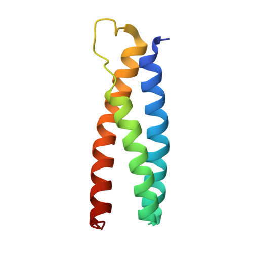
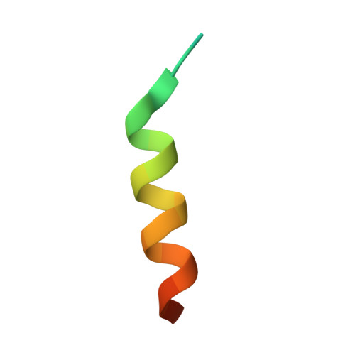
4U7I

Structure of the complex of Spartin MIT and IST1 MIM

  • Classification: PROTEIN TRANSPORT
  • Organism(s): Homo sapiens
  • Expression System: Escherichia coli
  • Mutation(s): No 

  • Deposited: 2014-07-30 Released: 2015-02-11 
  • Deposition Author(s): Guo, E.Z., Xu, Z.
  • Funding Organization(s): National Institutes of Health/National Institute of General Medical Sciences (NIH/NIGMS)

Experimental Data Snapshot

  • Method: X-RAY DIFFRACTION
  • Resolution: 1.79 Å
  • R-Value Free: 0.220 
  • R-Value Work: 0.190 
  • R-Value Observed: 0.192 

Starting Model: experimental
View more details

wwPDB Validation   3D Report Full Report


This is version 1.5 of the entry. See complete history


Literature

Distinct Mechanisms of Recognizing Endosomal Sorting Complex Required for Transport III (ESCRT-III) Protein IST1 by Different Microtubule Interacting and Trafficking (MIT) Domains.

Guo, E.Z.Xu, Z.

(2015) J Biol Chem 290: 8396-8408

  • DOI: https://doi.org/10.1074/jbc.M114.607903
  • Primary Citation of Related Structures:  
    4U7E, 4U7I, 4U7Y

  • PubMed Abstract: 

    The endosomal sorting complex required for transport (ESCRT) machinery is responsible for membrane remodeling in a number of biological processes including multivesicular body biogenesis, cytokinesis, and enveloped virus budding. In mammalian cells, efficient abscission during cytokinesis requires proper function of the ESCRT-III protein IST1, which binds to the microtubule interacting and trafficking (MIT) domains of VPS4, LIP5, and Spartin via its C-terminal MIT-interacting motif (MIM). Here, we studied the molecular interactions between IST1 and the three MIT domain-containing proteins to understand the structural basis that governs pairwise MIT-MIM interaction. Crystal structures of the three molecular complexes revealed that IST1 binds to the MIT domains of VPS4, LIP5, and Spartin using two different mechanisms (MIM1 mode versus MIM3 mode). Structural comparison revealed that structural features in both MIT and MIM contribute to determine the specific binding mechanism. Within the IST1 MIM sequence, two phenylalanine residues were shown to be important in discriminating MIM1 versus MIM3 binding. These observations enabled us to deduce a preliminary binding code, which we applied to provide CHMP2A, a protein that normally only binds the MIT domain in the MIM1 mode, the additional ability to bind the MIT domain of Spartin in the MIM3 mode.


  • Organizational Affiliation

    From the Life Science Institute and.


Macromolecules
Find similar proteins by:  (by identity cutoff)  |  3D Structure
Entity ID: 1
MoleculeChains Sequence LengthOrganismDetailsImage
Spartin95Homo sapiensMutation(s): 0 
Gene Names: SPG20KIAA0610TAHCCP1
UniProt & NIH Common Fund Data Resources
Find proteins for Q8N0X7 (Homo sapiens)
Explore Q8N0X7 
Go to UniProtKB:  Q8N0X7
PHAROS:  Q8N0X7
GTEx:  ENSG00000133104 
Entity Groups  
Sequence Clusters30% Identity50% Identity70% Identity90% Identity95% Identity100% Identity
UniProt GroupQ8N0X7
Sequence Annotations
Expand
  • Reference Sequence

Find similar proteins by:  Sequence   |   3D Structure  

Entity ID: 2
MoleculeChains Sequence LengthOrganismDetailsImage
IST1 homolog25Homo sapiensMutation(s): 0 
Gene Names: IST1KIAA0174
UniProt & NIH Common Fund Data Resources
Find proteins for P53990 (Homo sapiens)
Explore P53990 
Go to UniProtKB:  P53990
PHAROS:  P53990
GTEx:  ENSG00000182149 
Entity Groups  
Sequence Clusters30% Identity50% Identity70% Identity90% Identity95% Identity100% Identity
UniProt GroupP53990
Sequence Annotations
Expand
  • Reference Sequence
Experimental Data & Validation

Experimental Data

  • Method: X-RAY DIFFRACTION
  • Resolution: 1.79 Å
  • R-Value Free: 0.220 
  • R-Value Work: 0.190 
  • R-Value Observed: 0.192 
  • Space Group: P 43 21 2
Unit Cell:
Length ( Å )Angle ( ˚ )
a = 46.257α = 90
b = 46.257β = 90
c = 115.62γ = 90
Software Package:
Software NamePurpose
HKL-2000data scaling
PHASERphasing
PHENIXrefinement

Structure Validation

View Full Validation Report


Entry History & Funding Information

Deposition Data


Funding OrganizationLocationGrant Number
National Institutes of Health/National Institute of General Medical Sciences (NIH/NIGMS)United StatesGM095769

Revision History  (Full details and data files)

  • Version 1.0: 2015-02-11
    Type: Initial release
  • Version 1.1: 2015-02-18
    Changes: Database references
  • Version 1.2: 2015-04-08
    Changes: Database references
  • Version 1.3: 2017-09-27
    Changes: Author supporting evidence, Database references, Derived calculations, Other, Refinement description, Source and taxonomy
  • Version 1.4: 2019-12-25
    Changes: Author supporting evidence
  • Version 1.5: 2023-09-27
    Changes: Data collection, Database references, Refinement description