4U5W | pdb_00004u5w

Crystal Structure of HIV-1 Nef-SF2 Core Domain in Complex with the Src Family Kinase Hck SH3-SH2 Tandem Regulatory Domains


Experimental Data Snapshot

  • Method: X-RAY DIFFRACTION
  • Resolution: 1.86 Å
  • R-Value Free: 
    0.195 (Depositor), 0.192 (DCC) 
  • R-Value Work: 
    0.169 (Depositor), 0.173 (DCC) 
  • R-Value Observed: 
    0.170 (Depositor) 

Starting Models: experimental
View more details

wwPDB Validation 3D Report Full Report

Validation slider image for 4U5W

This is version 1.5 of the entry. See complete history

Literature

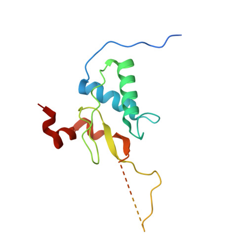
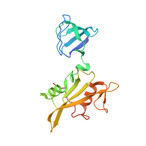
Interaction with the Src Homology (SH3-SH2) Region of the Src-family Kinase Hck Structures the HIV-1 Nef Dimer for Kinase Activation and Effector Recruitment.

Alvarado, J.J.Tarafdar, S.Yeh, J.I.Smithgall, T.E.

(2014) J Biological Chem 289: 28539-28553

  • DOI: https://doi.org/10.1074/jbc.M114.600031
  • Primary Citation Related Structures: 
    4U5W

  • PubMed Abstract: 

    HIV-1 Nef supports high titer viral replication in vivo and is essential for AIDS progression. Nef function depends on interactions with multiple host cell effectors, including Hck and other Src-family kinases. Here we describe the x-ray crystal structure of Nef in complex with the Hck SH3-SH2 regulatory region to a resolution of 1.86 Å. The complex crystallized as a dimer of complexes, with the conserved Nef PXXPXR motif engaging the Hck SH3 domain. A new intercomplex contact was found between SH3 Glu-93, and Nef Arg-105. Mutagenesis of Hck SH3 Glu-93 interfered with Nef·Hck complex formation and kinase activation in cells. The Hck SH2 domains impinge on the N-terminal region of Nef to stabilize a dimer conformation that exposes Asp-123, a residue critical for Nef function. Our results suggest that in addition to serving as a kinase effector for Nef, Hck binding may reorganize the Nef dimer for functional interaction with other signaling partners.


  • Organizational Affiliation
    • From the Departments of Microbiology and Molecular Genetics and Structural Biology, University of Pittsburgh School of Medicine, Pittsburgh, Pennsylvania 15219 and.

Macromolecule Content 

  • Total Structure Weight: 77.79 kDa 
  • Atom Count: 5,067 
  • Modeled Residue Count: 550 
  • Deposited Residue Count: 658 
  • Unique protein chains: 2

Macromolecules

Find similar proteins by:|  3D Structure
Entity ID: 1
MoleculeChains  Sequence LengthOrganismDetailsImage
Protein Nef
A, C
149HIV-1 M:B_ARV2/SF2Mutation(s): 0 
Gene Names: nef
UniProt
Find proteins for P03407 (Human immunodeficiency virus type 1 group M subtype B (isolate ARV2/SF2))
Explore P03407 
Go to UniProtKB:  P03407
Entity Groups
Sequence Clusters30% Identity50% Identity70% Identity90% Identity95% Identity100% Identity
UniProt GroupP03407
Sequence Annotations
Expand
Reference Sequence
Find similar proteins by:|  3D Structure
Entity ID: 2
MoleculeChains  Sequence LengthOrganismDetailsImage
Tyrosine-protein kinase HCK
B, D
180Homo sapiensMutation(s): 0 
Gene Names: HCK
EC: 2.7.10.2
UniProt & NIH Common Fund Data Resources
Find proteins for P08631 (Homo sapiens)
Explore P08631 
Go to UniProtKB:  P08631
PHAROS:  P08631
GTEx:  ENSG00000101336 
Entity Groups
Sequence Clusters30% Identity50% Identity70% Identity90% Identity95% Identity100% Identity
UniProt GroupP08631
Sequence Annotations
Expand
Reference Sequence

Small Molecules

Ligands 2 Unique
IDChains Name / Formula / InChI Key2D Diagram3D Interactions
IOD

Query on IOD


Download:Ideal Coordinates CCD File
H [auth A]
I [auth B]
J [auth B]
K [auth C]
L [auth D]
H [auth A],
I [auth B],
J [auth B],
K [auth C],
L [auth D],
M [auth D]
IODIDE ION
I
XMBWDFGMSWQBCA-UHFFFAOYSA-M
MPD

Query on MPD


Download:Ideal Coordinates CCD File
E [auth A],
F [auth A],
G [auth A]
(4S)-2-METHYL-2,4-PENTANEDIOL
C6 H14 O2
SVTBMSDMJJWYQN-YFKPBYRVSA-N

Experimental Data & Validation

Experimental Data

  • Method: X-RAY DIFFRACTION
  • Resolution: 1.86 Å
  • R-Value Free:  0.195 (Depositor), 0.192 (DCC) 
  • R-Value Work:  0.169 (Depositor), 0.173 (DCC) 
  • R-Value Observed: 0.170 (Depositor) 
Space Group: P 1
Unit Cell:
Length ( Å )Angle ( ˚ )
a = 56.489α = 112.28
b = 57.265β = 96.09
c = 72.111γ = 106
Software Package:
Software NamePurpose
PHENIXrefinement

Structure Validation

View Full Validation Report


Entry History 

& Funding Information

Deposition Data


Funding OrganizationLocationGrant Number
National Institutes of Health/National Institute Of Allergy and Infectious Diseases (NIH/NIAID)United StatesAI057083
National Institutes of Health/National Institute Of Allergy and Infectious Diseases (NIH/NIAID)United StatesAI102724
National Institutes of Health/National Institute of General Medical Sciences (NIH/NIGMS)United StatesGM082251

Revision History  (Full details and data files)

  • Version 1.0: 2014-08-20
    Type: Initial release
  • Version 1.1: 2014-08-27
    Changes: Database references
  • Version 1.2: 2014-10-22
    Changes: Database references
  • Version 1.3: 2017-09-13
    Changes: Advisory, Author supporting evidence, Database references, Derived calculations, Other, Source and taxonomy
  • Version 1.4: 2019-12-11
    Changes: Author supporting evidence
  • Version 1.5: 2023-09-27
    Changes: Data collection, Database references, Refinement description