


Crystal structure of the metallo-beta-lactamase NDM-1 in complex with a bisthiazolidine inhibitor
Kosmopoulou, M., Hinchliffe, P., Spencer, J.To be published.
Experimental Data Snapshot
Starting Model: experimental
View more details
Entity ID: 1 | |||||
|---|---|---|---|---|---|
| Molecule | Chains | Sequence Length | Organism | Details | Image |
| Beta-lactamase NDM-1 | 246 | Klebsiella pneumoniae | Mutation(s): 0 Gene Names: blaNDM-1 EC: 3.5.2.6 | 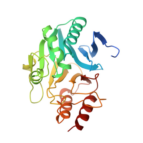 | |
UniProt | |||||
Entity Groups | |||||
| Sequence Clusters | 30% Identity50% Identity70% Identity90% Identity95% Identity100% Identity | ||||
| UniProt Group | C7C422 | ||||
Sequence AnnotationsExpand | |||||
Reference Sequence | |||||
| Ligands 3 Unique | |||||
|---|---|---|---|---|---|
| ID | Chains | Name / Formula / InChI Key | 2D Diagram | 3D Interactions | |
| 3C7 Download:Ideal Coordinates CCD File | G [auth A], J [auth B], N [auth C], R [auth D] | (3R,5R,7aS)-5-(sulfanylmethyl)tetrahydro[1,3]thiazolo[4,3-b][1,3]thiazole-3-carboxylic acid C7 H11 N O2 S3 ZTWVMVSSSBGFHH-JKUQZMGJSA-N |  | ||
| GOL Download:Ideal Coordinates CCD File | M [auth C], Q [auth D] | GLYCEROL C3 H8 O3 PEDCQBHIVMGVHV-UHFFFAOYSA-N |  | ||
| ZN Download:Ideal Coordinates CCD File | E [auth A] F [auth A] H [auth B] I [auth B] K [auth C] | ZINC ION Zn PTFCDOFLOPIGGS-UHFFFAOYSA-N |  | ||
| Length ( Å ) | Angle ( ˚ ) |
|---|---|
| a = 46.57 | α = 87.39 |
| b = 69.023 | β = 88.21 |
| c = 69.646 | γ = 76.75 |
| Software Name | Purpose |
|---|---|
| PHENIX | refinement |
| SCALA | data scaling |
| PHASER | phasing |
| Funding Organization | Location | Grant Number |
|---|---|---|
| National Institutes of Health | United States | AI100560 |


