The X-ray Structures of Six Octameric RNA Duplexes in the Presence of Different Di- and Trivalent Cations.
Schaffer, M.F., Peng, G., Spingler, B., Schnabl, J., Wang, M., Olieric, V., Sigel, R.K.(2016) Int J Mol Sci 17
- PubMed: 27355942 Search on PubMedSearch on PubMed Central
- DOI: https://doi.org/10.3390/ijms17070988
- Primary Citation Related Structures:
4U3L, 4U3O, 4U3P, 4U3R, 4U47, 4U78 - PubMed Abstract:
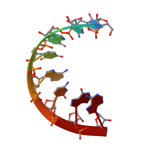
Due to the polyanionic nature of RNA, the principles of charge neutralization and electrostatic condensation require that cations help to overcome the repulsive forces in order for RNA to adopt a three-dimensional structure. A precise structural knowledge of RNA-metal ion interactions is crucial to understand the mechanism of metal ions in the catalytic or regulatory activity of RNA. We solved the crystal structure of an octameric RNA duplex in the presence of the di- and trivalent metal ions Ca(2+), Mn(2+), Co(2+), Cu(2+), Sr(2+), and Tb(3+). The detailed investigation reveals a unique innersphere interaction to uracil and extends the knowledge of the influence of metal ions for conformational changes in RNA structure. Furthermore, we could demonstrate that an accurate localization of the metal ions in the X-ray structures require the consideration of several crystallographic and geometrical parameters as well as the anomalous difference map.
- Department of Chemistry, University of Zurich, Winterthurerstrasse 190, Zürich CH-8057, Switzerland. michelle.schaffer@chem.uzh.ch.
Organizational Affiliation: