4U2Z

X-ray crystal structure of an Sco GlgEI-V279S/1,2,2-trifluromaltose complex


Experimental Data Snapshot

  • Method: X-RAY DIFFRACTION
  • Resolution: 2.26 Å
  • R-Value Free: 0.199 
  • R-Value Work: 0.163 
  • R-Value Observed: 0.165 

wwPDB Validation   3D Report Full Report


This is version 2.1 of the entry. See complete history


Literature

Synthesis of 2-deoxy-2,2-difluoro-alpha-maltosyl fluoride and its X-ray structure in complex with Streptomyces coelicolor GlgEI-V279S.

Thanna, S.Lindenberger, J.J.Gaitonde, V.V.Ronning, D.R.Sucheck, S.J.

(2015) Org Biomol Chem 13: 7542-7550

  • DOI: https://doi.org/10.1039/c5ob00867k
  • Primary Citation of Related Structures:  
    4U2Z

  • PubMed Abstract: 

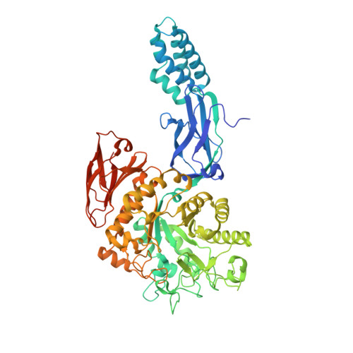
    Streptomyces coelicolor (Sco) GlgEI is a glycoside hydrolase involved in α-glucan biosynthesis and can be used as a model enzyme for structure-based inhibitor design targeting Mycobacterium tuberculosis (Mtb) GlgE. The latter is a genetically validated drug target for the development of anti-Tuberculosis (TB) treatments. Inhibition of Mtb GlgE results in a lethal buildup of the GlgE substrate maltose-1-phosphate (M1P). However, Mtb GlgE is difficult to crystallize and affords lower resolution X-ray structures. Sco GlgEI-V279S on the other hand crystallizes readily, produces high resolution X-ray data, and has active site topology identical to Mtb GlgE. We report the X-ray structure of Sco GlgEI-V279S in complex with 2-deoxy-2,2-difluoro-α-maltosyl fluoride (α-MTF, 5) at 2.3 Å resolution. α-MTF was designed as a non-hydrolysable mimic of M1P to probe the active site of GlgE1 prior to covalent bond formation without disruption of catalytic residues. The α-MTF complex revealed hydrogen bonding between Glu423 and the C1F which provides evidence that Glu423 functions as proton donor during catalysis. Further, hydrogen bonding between Arg392 and the axial C2 difluoromethylene moiety of α-MTF was observed suggesting that the C2 position tolerates substitution with hydrogen bond acceptors. The key step in the synthesis of α-MDF was transformation of peracetylated 2-fluoro-maltal 1 into peracetylated 2,2-difluoro-α-maltosyl fluoride 2 in a single step via the use of Selectfluor®.


  • Organizational Affiliation

    Department of Chemistry and Biochemistry, The University of Toledo, 2801 W. Bancroft Street, MS602, Toledo, OH, USA 43606. donald.ronning@utoledo.edu steve.sucheck@utoledo.edu.


Macromolecules
Find similar proteins by:  (by identity cutoff)  |  3D Structure
Entity ID: 1
MoleculeChains Sequence LengthOrganismDetailsImage
Alpha-1,4-glucan:maltose-1-phosphate maltosyltransferase 1
A, B
683Streptomyces coelicolor A3(2)Mutation(s): 1 
Gene Names: glgE1pep1pep1Apep1ISCO5443SC6A11.19c
EC: 2.4.99.16
UniProt
Find proteins for Q9L1K2 (Streptomyces coelicolor (strain ATCC BAA-471 / A3(2) / M145))
Explore Q9L1K2 
Go to UniProtKB:  Q9L1K2
Entity Groups  
Sequence Clusters30% Identity50% Identity70% Identity90% Identity95% Identity100% Identity
UniProt GroupQ9L1K2
Sequence Annotations
Expand
  • Reference Sequence
Oligosaccharides

Help

Entity ID: 2
MoleculeChains Length2D Diagram Glycosylation3D Interactions
alpha-D-glucopyranose-(1-4)-2-deoxy-2,2-difluoro-alpha-D-arabino-hexopyranosyl fluoride
C, D
2N/AN/A
Biologically Interesting Molecules (External Reference) 1 Unique
Experimental Data & Validation

Experimental Data

  • Method: X-RAY DIFFRACTION
  • Resolution: 2.26 Å
  • R-Value Free: 0.199 
  • R-Value Work: 0.163 
  • R-Value Observed: 0.165 
  • Space Group: P 41 21 2
Unit Cell:
Length ( Å )Angle ( ˚ )
a = 113.297α = 90
b = 113.297β = 90
c = 314.33γ = 90
Software Package:
Software NamePurpose
HKL-3000phasing
PDB_EXTRACTdata extraction
PHENIXrefinement

Structure Validation

View Full Validation Report


Entry History & Funding Information

Deposition Data


Funding OrganizationLocationGrant Number
National Institutes of Health/National Institute Of Allergy and Infectious Diseases (NIH/NIAID)United StatesAI105084

Revision History  (Full details and data files)

  • Version 1.0: 2015-07-22
    Type: Initial release
  • Version 1.1: 2015-08-12
    Changes: Database references
  • Version 1.2: 2017-09-20
    Changes: Author supporting evidence, Derived calculations, Refinement description
  • Version 1.3: 2019-12-11
    Changes: Author supporting evidence
  • Version 2.0: 2020-07-29
    Type: Remediation
    Reason: Carbohydrate remediation
    Changes: Atomic model, Data collection, Derived calculations, Non-polymer description, Structure summary
  • Version 2.1: 2023-12-27
    Changes: Data collection, Database references, Structure summary