4U0W

Crystal structure of YvoA from Bacillus subtilis in complex with N-acetylglucosamine-6-phosphate


Experimental Data Snapshot

  • Method: X-RAY DIFFRACTION
  • Resolution: 2.00 Å
  • R-Value Free: 0.254 
  • R-Value Work: 0.205 
  • R-Value Observed: 0.208 

wwPDB Validation   3D Report Full Report


Ligand Structure Quality Assessment 


This is version 2.1 of the entry. See complete history


Literature

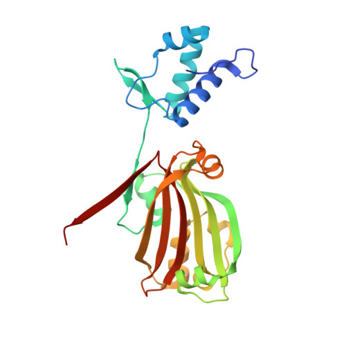
Structural insight into operator dre-sites recognition and effector binding in the GntR/HutC transcription regulator NagR.

Fillenberg, S.B.Grau, F.C.Seidel, G.Muller, Y.A.

(2015) Nucleic Acids Res 43: 1283-1296

  • DOI: https://doi.org/10.1093/nar/gku1374
  • Primary Citation of Related Structures:  
    4U0V, 4U0W, 4U0Y, 4WWC

  • PubMed Abstract: 

    The uptake and metabolism of N-acetylglucosamine (GlcNAc) in Bacillus subtilis is controlled by NagR (formerly named YvoA), a member of the widely-occurring GntR/HutC family of transcription regulators. Upon binding to specific DNA operator sites (dre-sites) NagR blocks the transcription of genes for GlcNAc utilization and interaction of NagR with effectors abrogates gene repression. Here we report crystal structures of NagR in complex with operator DNA and in complex with the putative effector molecules glucosamine-6-phosphate (GlcN-6-P) and N-acetylglucosamine-6-phosphate (GlcNAc-6-P). A comparison of the distinct conformational states suggests that effectors are able to displace the NagR-DNA-binding domains (NagR-DBDs) by almost 70 Å upon binding. In addition, a high-resolution crystal structure of isolated NagR-DBDs in complex with palindromic double-stranded DNA (dsDNA) discloses both the determinants for highly sequence-specific operator dre-site recognition and for the unspecific binding of NagR to dsDNA. Extensive biochemical binding studies investigating the affinities of full-length NagR and isolated NagR-DBDs for either random DNA, dre-site-derived palindromic or naturally occurring non-palindromic dre-site sequences suggest that proper NagR function relies on an effector-induced fine-tuning of the DNA-binding affinities of NagR and not on a complete abrogation of its DNA binding.


  • Organizational Affiliation

    Lehrstuhl für Biotechnik, Department of Biology, Friedrich-Alexander University Erlangen-Nuremberg, Henkestrasse 91, D-91052 Erlangen, Germany.


Macromolecules
Find similar proteins by:  (by identity cutoff)  |  3D Structure
Entity ID: 1
MoleculeChains Sequence LengthOrganismDetailsImage
HTH-type transcriptional repressor YvoA
A, B
246Bacillus subtilis subsp. subtilis str. 168Mutation(s): 0 
Gene Names: yvoABSU35030
UniProt
Find proteins for O34817 (Bacillus subtilis (strain 168))
Explore O34817 
Go to UniProtKB:  O34817
Entity Groups  
Sequence Clusters30% Identity50% Identity70% Identity90% Identity95% Identity100% Identity
UniProt GroupO34817
Sequence Annotations
Expand
  • Reference Sequence
Small Molecules
Ligands 3 Unique
IDChains Name / Formula / InChI Key2D Diagram3D Interactions
16G
Query on 16G

Download Ideal Coordinates CCD File 
C [auth A],
K [auth B]
2-acetamido-2-deoxy-6-O-phosphono-alpha-D-glucopyranose
C8 H16 N O9 P
BRGMHAYQAZFZDJ-PVFLNQBWSA-N
GOL
Query on GOL

Download Ideal Coordinates CCD File 
D [auth A],
L [auth B]
GLYCEROL
C3 H8 O3
PEDCQBHIVMGVHV-UHFFFAOYSA-N
EDO
Query on EDO

Download Ideal Coordinates CCD File 
E [auth A]
F [auth A]
G [auth A]
H [auth A]
I [auth A]
E [auth A],
F [auth A],
G [auth A],
H [auth A],
I [auth A],
J [auth A],
M [auth B],
N [auth B],
O [auth B],
P [auth B],
Q [auth B],
R [auth B],
S [auth B]
1,2-ETHANEDIOL
C2 H6 O2
LYCAIKOWRPUZTN-UHFFFAOYSA-N
Binding Affinity Annotations 
IDSourceBinding Affinity
16G Binding MOAD:  4U0W Kd: 1.00e+6 (nM) from 1 assay(s)
Experimental Data & Validation

Experimental Data

  • Method: X-RAY DIFFRACTION
  • Resolution: 2.00 Å
  • R-Value Free: 0.254 
  • R-Value Work: 0.205 
  • R-Value Observed: 0.208 
  • Space Group: H 3 2
Unit Cell:
Length ( Å )Angle ( ˚ )
a = 99.142α = 90
b = 99.142β = 90
c = 353.978γ = 120
Software Package:
Software NamePurpose
XSCALEdata scaling
PHASERphasing
PHENIXrefinement
XDSdata reduction

Structure Validation

View Full Validation Report


Ligand Structure Quality Assessment 


Entry History & Funding Information

Deposition Data


Funding OrganizationLocationGrant Number
German Research FoundationGermany--

Revision History  (Full details and data files)

  • Version 1.0: 2015-01-14
    Type: Initial release
  • Version 1.1: 2015-01-21
    Changes: Database references
  • Version 1.2: 2015-02-04
    Changes: Database references
  • Version 2.0: 2020-07-29
    Type: Remediation
    Reason: Carbohydrate remediation
    Changes: Advisory, Atomic model, Author supporting evidence, Data collection, Derived calculations, Structure summary
  • Version 2.1: 2023-12-20
    Changes: Data collection, Database references, Refinement description, Structure summary