4TYT

Crystal Structure of BcII metallo-beta-lactamase in complex with ML302F


Experimental Data Snapshot

  • Method: X-RAY DIFFRACTION
  • Resolution: 1.80 Å
  • R-Value Free: 0.201 
  • R-Value Work: 0.150 
  • R-Value Observed: 0.155 

Starting Model: experimental
View more details

wwPDB Validation   3D Report Full Report


Ligand Structure Quality Assessment 


This is version 3.0 of the entry. See complete history


Literature

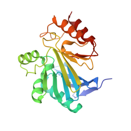
Rhodanine hydrolysis leads to potent thioenolate mediated metallo-beta-lactamase inhibition.

Brem, J.van Berkel, S.S.Aik, W.Rydzik, A.M.Avison, M.B.Pettinati, I.Umland, K.D.Kawamura, A.Spencer, J.Claridge, T.D.McDonough, M.A.Schofield, C.J.

(2014) Nat Chem 6: 1084-1090

  • DOI: https://doi.org/10.1038/nchem.2110
  • Primary Citation of Related Structures:  
    4PVO, 4PVT, 4TYT

  • PubMed Abstract: 

    The use of β-lactam antibiotics is compromised by resistance, which is provided by β-lactamases belonging to both metallo (MBL)- and serine (SBL)-β-lactamase subfamilies. The rhodanines are one of very few compound classes that inhibit penicillin-binding proteins (PBPs), SBLs and, as recently reported, MBLs. Here, we describe crystallographic analyses of the mechanism of inhibition of the clinically relevant VIM-2 MBL by a rhodanine, which reveal that the rhodanine ring undergoes hydrolysis to give a thioenolate. The thioenolate is found to bind via di-zinc chelation, mimicking the binding of intermediates in β-lactam hydrolysis. Crystallization of VIM-2 in the presence of the intact rhodanine led to observation of a ternary complex of MBL, a thioenolate fragment and rhodanine. The crystallographic observations are supported by kinetic and biophysical studies, including (19)F NMR analyses, which reveal the rhodanine-derived thioenolate to be a potent broad-spectrum MBL inhibitor and a lead structure for the development of new types of clinically useful MBL inhibitors.


  • Organizational Affiliation

    Department of Chemistry, University of Oxford, 12 Mansfield Road, Oxford OX1 3TA, UK.


Macromolecules
Find similar proteins by:  (by identity cutoff)  |  3D Structure
Entity ID: 1
MoleculeChains Sequence LengthOrganismDetailsImage
Beta-lactamase 2227Bacillus cereusMutation(s): 0 
Gene Names: blm
EC: 3.5.2.6
UniProt
Find proteins for P04190 (Bacillus cereus)
Explore P04190 
Go to UniProtKB:  P04190
Entity Groups  
Sequence Clusters30% Identity50% Identity70% Identity90% Identity95% Identity100% Identity
UniProt GroupP04190
Sequence Annotations
Expand
  • Reference Sequence
Experimental Data & Validation

Experimental Data

  • Method: X-RAY DIFFRACTION
  • Resolution: 1.80 Å
  • R-Value Free: 0.201 
  • R-Value Work: 0.150 
  • R-Value Observed: 0.155 
  • Space Group: C 1 2 1
Unit Cell:
Length ( Å )Angle ( ˚ )
a = 53.079α = 90
b = 61.358β = 93
c = 69.416γ = 90
Software Package:
Software NamePurpose
CrystalCleardata collection
DENZOdata reduction
PHENIXrefinement
Cootmodel building
SCALEPACKdata scaling
PHASERphasing

Structure Validation

View Full Validation Report


Ligand Structure Quality Assessment 


Entry History & Funding Information

Deposition Data


Funding OrganizationLocationGrant Number
Medical Research Council (United Kingdom)United KingdomG1100135

Revision History  (Full details and data files)

  • Version 1.0: 2014-11-26
    Type: Initial release
  • Version 1.1: 2014-12-03
    Changes: Database references
  • Version 2.0: 2017-09-13
    Changes: Advisory, Atomic model, Author supporting evidence, Derived calculations
  • Version 3.0: 2023-12-20
    Changes: Atomic model, Data collection, Database references, Refinement description