4TWP | pdb_00004twp

The crystal structure of human abl1 T315I gatekeeper mutant kinase domain in complex with axitinib


Experimental Data Snapshot

  • Method: X-RAY DIFFRACTION
  • Resolution: 2.40 Å
  • R-Value Free: 
    0.241 (Depositor), 0.237 (DCC) 
  • R-Value Work: 
    0.212 (Depositor), 0.206 (DCC) 
  • R-Value Observed: 
    0.213 (Depositor) 

wwPDB Validation 3D Report Full Report

Validation slider image for 4TWP

Ligand Structure Quality Assessment 


This is version 1.6 of the entry. See complete history

Literature

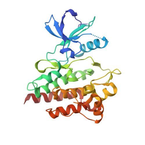
Axitinib effectively inhibits BCR-ABL1(T315I) with a distinct binding conformation.

Pemovska, T.Johnson, E.Kontro, M.Repasky, G.A.Chen, J.Wells, P.Cronin, C.N.McTigue, M.Kallioniemi, O.Porkka, K.Murray, B.W.Wennerberg, K.

(2015) Nature 519: 102-105

  • DOI: https://doi.org/10.1038/nature14119
  • Primary Citation Related Structures: 
    4TWP, 4WA9

  • PubMed Abstract: 

    The BCR-ABL1 fusion gene is a driver oncogene in chronic myeloid leukaemia and 30-50% of cases of adult acute lymphoblastic leukaemia. Introduction of ABL1 kinase inhibitors (for example, imatinib) has markedly improved patient survival, but acquired drug resistance remains a challenge. Point mutations in the ABL1 kinase domain weaken inhibitor binding and represent the most common clinical resistance mechanism. The BCR-ABL1 kinase domain gatekeeper mutation Thr315Ile (T315I) confers resistance to all approved ABL1 inhibitors except ponatinib, which has toxicity limitations. Here we combine comprehensive drug sensitivity and resistance profiling of patient cells ex vivo with structural analysis to establish the VEGFR tyrosine kinase inhibitor axitinib as a selective and effective inhibitor for T315I-mutant BCR-ABL1-driven leukaemia. Axitinib potently inhibited BCR-ABL1(T315I), at both biochemical and cellular levels, by binding to the active form of ABL1(T315I) in a mutation-selective binding mode. These findings suggest that the T315I mutation shifts the conformational equilibrium of the kinase in favour of an active (DFG-in) A-loop conformation, which has more optimal binding interactions with axitinib. Treatment of a T315I chronic myeloid leukaemia patient with axitinib resulted in a rapid reduction of T315I-positive cells from bone marrow. Taken together, our findings demonstrate an unexpected opportunity to repurpose axitinib, an anti-angiogenic drug approved for renal cancer, as an inhibitor for ABL1 gatekeeper mutant drug-resistant leukaemia patients. This study shows that wild-type proteins do not always sample the conformations available to disease-relevant mutant proteins and that comprehensive drug testing of patient-derived cells can identify unpredictable, clinically significant drug-repositioning opportunities.


  • Organizational Affiliation
    • Institute for Molecular Medicine Finland (FIMM), University of Helsinki, 00290 Helsinki, Finland.

Macromolecule Content 

  • Total Structure Weight: 63.91 kDa 
  • Atom Count: 4,617 
  • Modeled Residue Count: 537 
  • Deposited Residue Count: 542 
  • Unique protein chains: 1

Macromolecules

Find similar proteins by:|  3D Structure
Entity ID: 1
MoleculeChains  Sequence LengthOrganismDetailsImage
Tyrosine-protein kinase ABL1
A, B
271Homo sapiensMutation(s): 1 
Gene Names: ABL1ABLJTK7
EC: 2.7.10.2
UniProt & NIH Common Fund Data Resources
Find proteins for P00519 (Homo sapiens)
Explore P00519 
Go to UniProtKB:  P00519
PHAROS:  P00519
GTEx:  ENSG00000097007 
Entity Groups
Sequence Clusters30% Identity50% Identity70% Identity90% Identity95% Identity100% Identity
UniProt GroupP00519
Sequence Annotations
Expand
Reference Sequence

Small Molecules

Ligands 3 Unique
IDChains Name / Formula / InChI Key2D Diagram3D Interactions
AXI

Query on AXI


Download:Ideal Coordinates CCD File
C [auth A],
H [auth B]
AXITINIB
C22 H18 N4 O S
RITAVMQDGBJQJZ-FMIVXFBMSA-N
NI

Query on NI


Download:Ideal Coordinates CCD File
D [auth A],
E [auth A],
I [auth B],
J [auth B]
NICKEL (II) ION
Ni
VEQPNABPJHWNSG-UHFFFAOYSA-N
NA

Query on NA


Download:Ideal Coordinates CCD File
F [auth A],
G [auth A],
K [auth B]
SODIUM ION
Na
FKNQFGJONOIPTF-UHFFFAOYSA-N
Binding Affinity Annotations 
IDSourceBinding Affinity
AXI BindingDB:  4TWP Kd: min: 1.5, max: 2600 (nM) from 15 assay(s)
IC50: min: 0.2, max: 170 (nM) from 8 assay(s)

Experimental Data & Validation

Experimental Data

  • Method: X-RAY DIFFRACTION
  • Resolution: 2.40 Å
  • R-Value Free:  0.241 (Depositor), 0.237 (DCC) 
  • R-Value Work:  0.212 (Depositor), 0.206 (DCC) 
  • R-Value Observed: 0.213 (Depositor) 
Space Group: P 2 21 21
Unit Cell:
Length ( Å )Angle ( ˚ )
a = 56.818α = 90
b = 113.801β = 90
c = 131.164γ = 90
Software Package:
Software NamePurpose
PHENIXrefinement
SCALAdata scaling
Cootmodel building
PHASERphasing
PDB_EXTRACTdata extraction
XSCALEdata scaling

Structure Validation

View Full Validation Report


Ligand Structure Quality Assessment 


Entry History 

Deposition Data

Revision History  (Full details and data files)

  • Version 1.0: 2015-02-11
    Type: Initial release
  • Version 1.1: 2015-03-04
    Changes: Database references
  • Version 1.2: 2015-03-18
    Changes: Database references
  • Version 1.3: 2015-05-13
    Changes: Database references
  • Version 1.4: 2017-11-22
    Changes: Derived calculations, Refinement description, Source and taxonomy
  • Version 1.5: 2023-12-27
    Changes: Data collection, Database references, Derived calculations
  • Version 1.6: 2024-08-07
    Changes: Data collection