4TWB

Sulfolobus solfataricus ribose-phosphate pyrophosphokinase


Experimental Data Snapshot

  • Method: X-RAY DIFFRACTION
  • Resolution: 2.80 Å
  • R-Value Free: 0.265 
  • R-Value Work: 0.235 
  • R-Value Observed: 0.236 

wwPDB Validation   3D Report Full Report


Ligand Structure Quality Assessment 


This is version 1.5 of the entry. See complete history


Literature

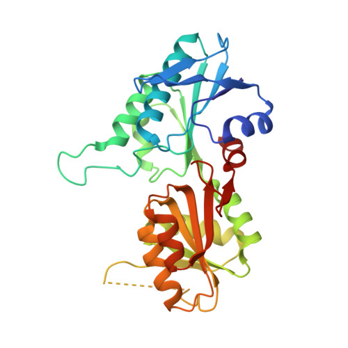
Structure of dimeric, recombinant Sulfolobus solfataricus phosphoribosyl diphosphate synthase: a bent dimer defining the adenine specificity of the substrate ATP.

Andersen, R.W.Leggio, L.L.Hove-Jensen, B.Kadziola, A.

(2015) Extremophiles 19: 407-415

  • DOI: https://doi.org/10.1007/s00792-014-0726-x
  • Primary Citation of Related Structures:  
    4TWB

  • PubMed Abstract: 

    The enzyme 5-phosphoribosyl-1-α-diphosphate (PRPP) synthase (EC 2.7.6.1) catalyses the Mg(2+)-dependent transfer of a diphosphoryl group from ATP to the C1 hydroxyl group of ribose 5-phosphate resulting in the production of PRPP and AMP. A nucleotide sequence specifying Sulfolobus solfataricus PRPP synthase was synthesised in vitro with optimised codon usage for expression in Escherichia coli. Following expression of the gene in E. coli PRPP synthase was purified by heat treatment and ammonium sulphate precipitation and the structure of S. solfataricus PRPP synthase was determined at 2.8 Å resolution. A bent dimer oligomerisation was revealed, which seems to be an abundant feature among PRPP synthases for defining the adenine specificity of the substrate ATP. Molecular replacement was used to determine the S. solfataricus PRPP synthase structure with a monomer subunit of Methanocaldococcus jannaschii PRPP synthase as a search model. The two amino acid sequences share 35 % identity. The resulting asymmetric unit consists of three separated dimers. The protein was co-crystallised in the presence of AMP and ribose 5-phosphate, but in the electron density map of the active site only AMP and a sulphate ion were observed. Sulphate ion, reminiscent of the ammonium sulphate precipitation step of the purification, seems to bind tightly and, therefore, presumably occupies and blocks the ribose 5-phosphate binding site. The activity of S. solfataricus PRPP synthase is independent of phosphate ion.


  • Organizational Affiliation

    Department of Chemistry, University of Copenhagen, 5 Universitetsparken, 2100, Copenhagen Ø, Denmark.


Macromolecules
Find similar proteins by:  (by identity cutoff)  |  3D Structure
Entity ID: 1
MoleculeChains Sequence LengthOrganismDetailsImage
Ribose-phosphate pyrophosphokinase
A, B, C, D, E
A, B, C, D, E, F
291Saccharolobus solfataricusMutation(s): 0 
EC: 2.7.6.1
UniProt
Find proteins for Q97Z86 (Saccharolobus solfataricus (strain ATCC 35092 / DSM 1617 / JCM 11322 / P2))
Explore Q97Z86 
Go to UniProtKB:  Q97Z86
Entity Groups  
Sequence Clusters30% Identity50% Identity70% Identity90% Identity95% Identity100% Identity
UniProt GroupQ97Z86
Sequence Annotations
Expand
  • Reference Sequence
Small Molecules
Ligands 2 Unique
IDChains Name / Formula / InChI Key2D Diagram3D Interactions
AMP
Query on AMP

Download Ideal Coordinates CCD File 
G [auth A]
I [auth A]
K [auth C]
M [auth C]
P [auth E]
G [auth A],
I [auth A],
K [auth C],
M [auth C],
P [auth E],
Q [auth F]
ADENOSINE MONOPHOSPHATE
C10 H14 N5 O7 P
UDMBCSSLTHHNCD-KQYNXXCUSA-N
SO4
Query on SO4

Download Ideal Coordinates CCD File 
H [auth A]
J [auth B]
L [auth C]
N [auth D]
O [auth E]
H [auth A],
J [auth B],
L [auth C],
N [auth D],
O [auth E],
R [auth F]
SULFATE ION
O4 S
QAOWNCQODCNURD-UHFFFAOYSA-L
Experimental Data & Validation

Experimental Data

  • Method: X-RAY DIFFRACTION
  • Resolution: 2.80 Å
  • R-Value Free: 0.265 
  • R-Value Work: 0.235 
  • R-Value Observed: 0.236 
  • Space Group: P 1 21 1
Unit Cell:
Length ( Å )Angle ( ˚ )
a = 80.217α = 90
b = 92.846β = 90.91
c = 138.338γ = 90
Software Package:
Software NamePurpose
SCALEPACKdata scaling
PHENIXrefinement

Structure Validation

View Full Validation Report


Ligand Structure Quality Assessment 


Entry History 

Deposition Data

  • Released Date: 2014-07-30 
  • Deposition Author(s): Kadziola, A.

Revision History  (Full details and data files)

  • Version 1.0: 2014-07-30
    Type: Initial release
  • Version 1.1: 2015-02-04
    Changes: Database references
  • Version 1.2: 2015-03-04
    Changes: Database references
  • Version 1.3: 2018-01-17
    Changes: Data collection
  • Version 1.4: 2018-02-14
    Changes: Data collection
  • Version 1.5: 2023-12-20
    Changes: Data collection, Database references, Refinement description