Computational design of a red fluorophore ligase for site-specific protein labeling in living cells.
Liu, D.S., Nivon, L.G., Richter, F., Goldman, P.J., Deerinck, T.J., Yao, J.Z., Richardson, D., Phipps, W.S., Ye, A.Z., Ellisman, M.H., Drennan, C.L., Baker, D., Ting, A.Y.(2014) Proc Natl Acad Sci U S A 111: E4551-E4559
- PubMed: 25313043 Search on PubMedSearch on PubMed Central
- DOI: https://doi.org/10.1073/pnas.1404736111
- Primary Citation Related Structures:
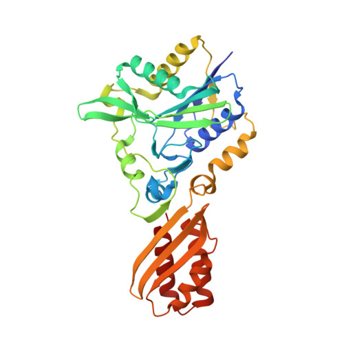
4TVW, 4TVY - PubMed Abstract:
Chemical fluorophores offer tremendous size and photophysical advantages over fluorescent proteins but are much more challenging to target to specific cellular proteins. Here, we used Rosetta-based computation to design a fluorophore ligase that accepts the red dye resorufin, starting from Escherichia coli lipoic acid ligase. X-ray crystallography showed that the design closely matched the experimental structure. Resorufin ligase catalyzed the site-specific and covalent attachment of resorufin to various cellular proteins genetically fused to a 13-aa recognition peptide in multiple mammalian cell lines and in primary cultured neurons. We used resorufin ligase to perform superresolution imaging of the intermediate filament protein vimentin by stimulated emission depletion and electron microscopies. This work illustrates the power of Rosetta for major redesign of enzyme specificity and introduces a tool for minimally invasive, highly specific imaging of cellular proteins by both conventional and superresolution microscopies.
- Departments of Chemistry and.
Organizational Affiliation: