4TT9 | pdb_00004tt9

Structure of the C-terminal SpoA domain of Shigella flexneri Spa33


Experimental Data Snapshot

  • Method: X-RAY DIFFRACTION
  • Resolution: 2.30 Å
  • R-Value Free: 
    0.210 (Depositor), 0.182 (DCC) 
  • R-Value Work: 
    0.174 (Depositor), 0.175 (DCC) 
  • R-Value Observed: 
    0.177 (Depositor) 

Starting Models: experimental
View more details

wwPDB Validation 3D Report Full Report

Validation slider image for 4TT9

This is version 1.3 of the entry. See complete history

Literature

Characterisation of Shigella Spa33 and Thermotoga FliM/N reveals a new model for C-ring assembly in T3SS.

McDowell, M.A.Marcoux, J.McVicker, G.Johnson, S.Fong, Y.H.Stevens, R.Bowman, L.A.Degiacomi, M.T.Yan, J.Wise, A.Friede, M.E.Benesch, J.L.Deane, J.E.Tang, C.M.Robinson, C.V.Lea, S.M.

(2016) Mol Microbiol 99: 749-766

  • DOI: https://doi.org/10.1111/mmi.13267
  • Primary Citation Related Structures: 
    4TT9

  • PubMed Abstract: 

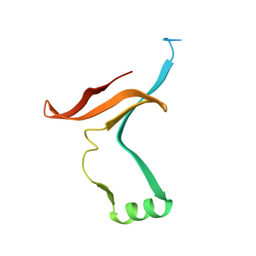
    Flagellar type III secretion systems (T3SS) contain an essential cytoplasmic-ring (C-ring) largely composed of two proteins FliM and FliN, whereas an analogous substructure for the closely related non-flagellar (NF) T3SS has not been observed in situ. We show that the spa33 gene encoding the putative NF-T3SS C-ring component in Shigella flexneri is alternatively translated to produce both full-length (Spa33-FL) and a short variant (Spa33-C), with both required for secretion. They associate in a 1:2 complex (Spa33-FL/C2) that further oligomerises into elongated arrays in vitro. The structure of Spa33-C2 and identification of an unexpected intramolecular pseudodimer in Spa33-FL reveal a molecular model for their higher order assembly within NF-T3SS. Spa33-FL and Spa33-C are identified as functional counterparts of a FliM-FliN fusion and free FliN respectively. Furthermore, we show that Thermotoga maritima FliM and FliN form a 1:3 complex structurally equivalent to Spa33-FL/C2 , allowing us to propose a unified model for C-ring assembly by NF-T3SS and flagellar-T3SS.


  • Organizational Affiliation
    • Sir William Dunn School of Pathology, University of Oxford, Oxford, UK.

Macromolecule Content 

  • Total Structure Weight: 39.79 kDa 
  • Atom Count: 2,354 
  • Modeled Residue Count: 282 
  • Deposited Residue Count: 344 
  • Unique protein chains: 1

Macromolecules

Find similar proteins by:|  3D Structure
Entity ID: 1
MoleculeChains  Sequence LengthOrganismDetailsImage
Surface presentation of antigens protein SpaO
A, B, C, D
86Shigella flexneriMutation(s): 0 
Gene Names: spaOspa33CP0152
UniProt
Find proteins for P0A1K9 (Shigella flexneri)
Explore P0A1K9 
Go to UniProtKB:  P0A1K9
Entity Groups
Sequence Clusters30% Identity50% Identity70% Identity90% Identity95% Identity100% Identity
UniProt GroupP0A1K9
Sequence Annotations
Expand
Reference Sequence

Experimental Data & Validation

Experimental Data

  • Method: X-RAY DIFFRACTION
  • Resolution: 2.30 Å
  • R-Value Free:  0.210 (Depositor), 0.182 (DCC) 
  • R-Value Work:  0.174 (Depositor), 0.175 (DCC) 
  • R-Value Observed: 0.177 (Depositor) 
Space Group: P 1 21 1
Unit Cell:
Length ( Å )Angle ( ˚ )
a = 51.3α = 90
b = 27.82β = 90.03
c = 104.44γ = 90
Software Package:
Software NamePurpose
PHENIXrefinement
XDSdata reduction
XSCALEdata scaling
Aimlessdata scaling
PHASERphasing

Structure Validation

View Full Validation Report


Entry History 

Deposition Data

Revision History  (Full details and data files)

  • Version 1.0: 2015-06-24
    Type: Initial release
  • Version 1.1: 2015-11-18
    Changes: Database references
  • Version 1.2: 2016-02-24
    Changes: Database references
  • Version 1.3: 2023-12-20
    Changes: Data collection, Database references, Refinement description