Differential Modes of Peptide Binding onto Replicative Sliding Clamps from Various Bacterial Origins.
Wolff, P., Amal, I., Olieric, V., Chaloin, O., Gygli, G., Ennifar, E., Lorber, B., Guichard, G., Wagner, J., Dejaegere, A., Burnouf, D.Y.(2014) J Med Chem 57: 7565-7576
- PubMed: 25170813 Search on PubMed
- DOI: https://doi.org/10.1021/jm500467a
- Primary Citation Related Structures:
4TR6, 4TR7, 4TR8, 4TSZ - PubMed Abstract:
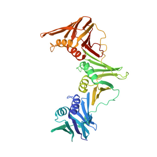
Bacterial sliding clamps are molecular hubs that interact with many proteins involved in DNA metabolism through their binding, via a conserved peptidic sequence, into a universally conserved pocket. This interacting pocket is acknowledged as a potential molecular target for the development of new antibiotics. We previously designed short peptides with an improved affinity for the Escherichia coli binding pocket. Here we show that these peptides differentially interact with other bacterial clamps, despite the fact that all pockets are structurally similar. Thermodynamic and modeling analyses of the interactions differentiate between two categories of clamps: group I clamps interact efficiently with our designed peptides and assemble the Escherichia coli and related orthologs clamps, whereas group II clamps poorly interact with the same peptides and include Bacillus subtilis and other Gram-positive clamps. These studies also suggest that the peptide binding process could occur via different mechanisms, which depend on the type of clamp.
- Université de Strasbourg , UPR9002, Architecture et Réactivité de l'ARN, Institut de Biologie Moléculaire et Cellulaire du CNRS, 15, rue René Descartes, 67084 Strasbourg, France.
Organizational Affiliation: