4TPI

THE REFINED 2.2-ANGSTROMS (0.22-NM) X-RAY CRYSTAL STRUCTURE OF THE TERNARY COMPLEX FORMED BY BOVINE TRYPSINOGEN, VALINE-VALINE AND THE ARG15 ANALOGUE OF BOVINE PANCREATIC TRYPSIN INHIBITOR


Experimental Data Snapshot

  • Method: X-RAY DIFFRACTION
  • Resolution: 2.20 Å
  • R-Value Work: 0.170 

wwPDB Validation   3D Report Full Report


This is version 1.2 of the entry. See complete history


Literature

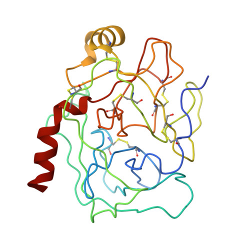
The refined 2.2-A (0.22-nm) X-ray crystal structure of the ternary complex formed by bovine trypsinogen, valine-valine and the Arg15 analogue of bovine pancreatic trypsin inhibitor

Bode, W.Walter, J.Huber, R.Wenzel, H.R.Tschesche, H.

(1984) Eur J Biochem 144: 185-190

  • DOI: https://doi.org/10.1111/j.1432-1033.1984.tb08447.x
  • Primary Citation of Related Structures:  
    4TPI

  • PubMed Abstract: 
  • Large orthorhombic crystals of the complex formed by bovine trypsinogen and a semisynthetic homologous bovine pancreatic trypsin inhibitor with the reactive-site lysine residue replaced by an arginine residue [( Arg15]PTI) have been obtained which are isomorphous with the crystals of PTI-trypsinogen [Bode, W ...

    Large orthorhombic crystals of the complex formed by bovine trypsinogen and a semisynthetic homologous bovine pancreatic trypsin inhibitor with the reactive-site lysine residue replaced by an arginine residue [( Arg15]PTI) have been obtained which are isomorphous with the crystals of PTI-trypsinogen [Bode, W., Schwager, P. and Huber, R. (1978) J. Mol. Biol. 118, 99-112]. The X-ray crystal structure of the ternary complex of trypsinogen-[Arg15]PTI with the dipeptide Val-Val has been determined by X-ray data to 2.2-A (0.22-nm) resolution by means of difference Fourier methods and has been crystallographically refined to a final R-value of 0.17. Replacement of the reactive-site Lys15 by an arginine residue is accompanied in the complex by small movements of polar side groups of trypsin and enclosed solvent molecules within the specificity pocket. Only solvent molecule 414 OH which mediates the hydrogen bond interactions between Lys15 NZ and Asp189 carboxylate is expelled, thus allowing the bulkier guanidyl group to approach this carboxylate. The dipeptide Val-Val binds in the pocket accepting the Ile-Val N-terminus in trypsin. The cavity left by the CD-methyl group of Ile16 upon replacement by a valine residue is only partially filled by slight rearrangements of neighbouring peptide side chains. Part of the positive free energy change observed upon replacement of Ile-Val may allow for the maintenance of this cavity.


    Related Citations: 
    • The Transition of Bovine Trypsinogen to a Trypsin-Like State Upon Strong Ligand Binding. II. The Binding of the Pancreatic Trypsin Inhibitor and of Isoleucine-Valine and of Sequentially Related Peptides to Trypsinogen and to P-Guanidinobenzoate-Trypsinogen
      Bode, W.
      (1979) J Mol Biol 127: 357
    • The Transition of Bovine Trypsinogen to a Trypsin-Like State Upon Strong Ligand Binding. The Refined Crystal Structures of the Bovine Trypsinogen-Pancreatic Trypsin Inhibitor Complex and of its Ternary Complex with Ile-Val at 1.9 Angstroms Resolution
      Bode, W., Schwager, P., Huber, R.
      (1978) J Mol Biol 118: 99

    Organizational Affiliation

    Department of Cell Biology, La Jolla Institute for Allergy and Immunology, La Jolla CA 92037, USA.


Macromolecules
Find similar proteins by:  (by identity cutoff)  |  3D Structure
Entity ID: 1
MoleculeChainsSequence LengthOrganismDetailsImage
TRYPSINOGENA [auth Z]229Bos taurusMutation(s): 0 
Gene Names: PRSS1TRP1TRY1TRYP1
EC: 3.4.21.4
UniProt
Find proteins for P00760 (Bos taurus)
Explore P00760 
Go to UniProtKB:  P00760
Entity Groups  
Sequence Clusters30% Identity50% Identity70% Identity90% Identity95% Identity100% Identity
UniProt GroupP00760
Protein Feature View
Expand
  • Reference Sequence
Find similar proteins by:  (by identity cutoff)  |  3D Structure
Entity ID: 2
MoleculeChainsSequence LengthOrganismDetailsImage
BOVINE PANCREATIC TRYPSIN INHIBITORB [auth I]58N/AMutation(s): 0 
UniProt
Find proteins for P00974 (Bos taurus)
Explore P00974 
Go to UniProtKB:  P00974
Entity Groups  
Sequence Clusters30% Identity50% Identity70% Identity90% Identity95% Identity100% Identity
UniProt GroupP00974
Protein Feature View
Expand
  • Reference Sequence
Small Molecules
Ligands 3 Unique
IDChainsName / Formula / InChI Key2D Diagram3D Interactions
VAL
Query on VAL

Download Ideal Coordinates CCD File 
C [auth Z],
D [auth Z]
VALINE
C5 H11 N O2
KZSNJWFQEVHDMF-BYPYZUCNSA-N
 Ligand Interaction
SO4
Query on SO4

Download Ideal Coordinates CCD File 
F [auth I],
G [auth I]
SULFATE ION
O4 S
QAOWNCQODCNURD-UHFFFAOYSA-L
 Ligand Interaction
CA
Query on CA

Download Ideal Coordinates CCD File 
E [auth Z]CALCIUM ION
Ca
BHPQYMZQTOCNFJ-UHFFFAOYSA-N
 Ligand Interaction
Experimental Data & Validation

Experimental Data

  • Method: X-RAY DIFFRACTION
  • Resolution: 2.20 Å
  • R-Value Work: 0.170 
  • Space Group: I 2 2 2
Unit Cell:
Length ( Å )Angle ( ˚ )
a = 75.51α = 90
b = 85.45β = 90
c = 122.3γ = 90
Software Package:
Software NamePurpose
EREFrefinement

Structure Validation

View Full Validation Report


Entry History 

Deposition Data

Revision History  (Full details and data files)

  • Version 1.0: 1985-11-08
    Type: Initial release
  • Version 1.1: 2008-03-25
    Changes: Version format compliance
  • Version 1.2: 2011-07-13
    Changes: Version format compliance