4TMU

Crystal structure of RecQ catalytic core from C. sakazakii bound to DNA


Experimental Data Snapshot

  • Method: X-RAY DIFFRACTION
  • Resolution: 2.40 Å
  • R-Value Free: 0.233 
  • R-Value Work: 0.179 
  • R-Value Observed: 0.181 

wwPDB Validation   3D Report Full Report


This is version 1.5 of the entry. See complete history


Literature

Structural mechanisms of DNA binding and unwinding in bacterial RecQ helicases.

Manthei, K.A.Hill, M.C.Burke, J.E.Butcher, S.E.Keck, J.L.

(2015) Proc Natl Acad Sci U S A 112: 4292-4297

  • DOI: https://doi.org/10.1073/pnas.1416746112
  • Primary Citation of Related Structures:  
    4TMU

  • PubMed Abstract: 

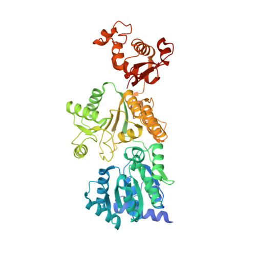
    RecQ helicases unwind remarkably diverse DNA structures as key components of many cellular processes. How RecQ enzymes accommodate different substrates in a unified mechanism that couples ATP hydrolysis to DNA unwinding is unknown. Here, the X-ray crystal structure of the Cronobacter sakazakii RecQ catalytic core domain bound to duplex DNA with a 3' single-stranded extension identifies two DNA-dependent conformational rearrangements: a winged-helix domain pivots ∼90° to close onto duplex DNA, and a conserved aromatic-rich loop is remodeled to bind ssDNA. These changes coincide with a restructuring of the RecQ ATPase active site that positions catalytic residues for ATP hydrolysis. Complex formation also induces a tight bend in the DNA and melts a portion of the duplex. This bending, coupled with translocation, could provide RecQ with a mechanism for unwinding duplex and other DNA structures.


  • Organizational Affiliation

    Department of Biomolecular Chemistry, University of Wisconsin School of Medicine and Public Health, Madison, WI 53706; and.


Macromolecules

Find similar proteins by:  (by identity cutoff)  |  3D Structure
Entity ID: 1
MoleculeChains Sequence LengthOrganismDetailsImage
RecQ541Cronobacter sakazakii ATCC BAA-894Mutation(s): 0 
Gene Names: ESA_03738
UniProt
Find proteins for A7MQK9 (Cronobacter sakazakii (strain ATCC BAA-894))
Explore A7MQK9 
Go to UniProtKB:  A7MQK9
Entity Groups  
Sequence Clusters30% Identity50% Identity70% Identity90% Identity95% Identity100% Identity
UniProt GroupA7MQK9
Sequence Annotations
Expand
  • Reference Sequence
Find similar nucleic acids by:  (by identity cutoff)  |  3D Structure
Entity ID: 2
MoleculeChains LengthOrganismImage
DNA (29-MER)34synthetic construct
Sequence Annotations
Expand
  • Reference Sequence
Small Molecules
Ligands 1 Unique
IDChains Name / Formula / InChI Key2D Diagram3D Interactions
ZN
Query on ZN

Download Ideal Coordinates CCD File 
C [auth A]ZINC ION
Zn
PTFCDOFLOPIGGS-UHFFFAOYSA-N
Experimental Data & Validation

Experimental Data

  • Method: X-RAY DIFFRACTION
  • Resolution: 2.40 Å
  • R-Value Free: 0.233 
  • R-Value Work: 0.179 
  • R-Value Observed: 0.181 
  • Space Group: P 21 21 2
Unit Cell:
Length ( Å )Angle ( ˚ )
a = 78.348α = 90
b = 93.959β = 90
c = 100.16γ = 90
Software Package:
Software NamePurpose
REFMACrefinement

Structure Validation

View Full Validation Report


Entry History & Funding Information

Deposition Data


Funding OrganizationLocationGrant Number
National Institutes of Health/National Institute of General Medical Sciences (NIH/NIGMS)United StatesP41GM103399
Department of Energy (DOE, United States)United StatesDE-AC02-06CH11357
National Institutes of Health/National Institute of General Medical Sciences (NIH/NIGMS)United StatesGM098885

Revision History  (Full details and data files)

  • Version 1.0: 2015-03-11
    Type: Initial release
  • Version 1.1: 2015-03-25
    Changes: Database references
  • Version 1.2: 2015-04-15
    Changes: Database references
  • Version 1.3: 2015-04-22
    Changes: Database references
  • Version 1.4: 2020-01-01
    Changes: Author supporting evidence, Database references, Derived calculations, Other, Source and taxonomy
  • Version 1.5: 2023-12-27
    Changes: Data collection, Database references, Refinement description