4TKY

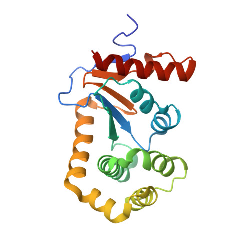
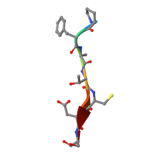
The complex structure of E. coli DsbA bound to a peptide at the DsbA/DsbB interface


Experimental Data Snapshot

  • Method: X-RAY DIFFRACTION
  • Resolution: 2.50 Å
  • R-Value Free: 0.237 
  • R-Value Work: 0.176 
  • R-Value Observed: 0.179 

Starting Model: experimental
View more details

wwPDB Validation   3D Report Full Report


This is version 1.4 of the entry. See complete history


Literature

Peptide Inhibitors of the Escherichia coli DsbA Oxidative Machinery Essential for Bacterial Virulence.

Duprez, W.Premkumar, L.Halili, M.A.Lindahl, F.Reid, R.C.Fairlie, D.P.Martin, J.L.

(2015) J Med Chem 58: 577-587

  • DOI: https://doi.org/10.1021/jm500955s
  • Primary Citation of Related Structures:  
    4TKY

  • PubMed Abstract: 

    One approach to address antibiotic resistance is to develop drugs that interfere with bacterial virulence. A master regulator of virulence in Gram-negative bacteria is the oxidative folding machinery comprising DsbA and DsbB. A crystal structure at 2.5 Å resolution is reported here for Escherichia coli DsbA complexed with PFATCDS, a heptapeptide derived from the partner protein Escherichia coli DsbB. Details of the peptide binding mode and binding site provide valuable clues for inhibitor design. Structure-activity relationships for 30 analogues were used to produce short peptides with a cysteine that bind tightly to EcDsbA (Kd = 2.0 ± 0.3 μM) and inhibit its activity (IC50 = 5.1 ± 1.1 μM). The most potent inhibitor does not bind to or inhibit human thioredoxin that shares a similar active site. This finding suggests that small molecule inhibitors can be designed to exploit a key interaction of EcDsbA, as the basis for antivirulence agents with a novel mechanism of action.


  • Organizational Affiliation

    Institute for Molecular Bioscience, Division of Chemistry and Structural Biology, University of Queensland , Brisbane, Queensland 4072, Australia.


Macromolecules
Find similar proteins by:  (by identity cutoff)  |  3D Structure
Entity ID: 1
MoleculeChains Sequence LengthOrganismDetailsImage
Thiol:disulfide interchange protein DsbA
A, B, C, D
191Escherichia coli K-12Mutation(s): 0 
Gene Names: dsbAdsfppfAb3860JW3832
UniProt
Find proteins for P0AEG4 (Escherichia coli (strain K12))
Explore P0AEG4 
Go to UniProtKB:  P0AEG4
Entity Groups  
Sequence Clusters30% Identity50% Identity70% Identity90% Identity95% Identity100% Identity
UniProt GroupP0AEG4
Sequence Annotations
Expand
  • Reference Sequence

Find similar proteins by:  Sequence   |   3D Structure  

Entity ID: 2
MoleculeChains Sequence LengthOrganismDetailsImage
PRO-PHE-ALA-THR-CYS-ASP-SERE [auth F],
F [auth E],
G,
H
7Escherichia coliMutation(s): 0 
Sequence Annotations
Expand
  • Reference Sequence
Experimental Data & Validation

Experimental Data

  • Method: X-RAY DIFFRACTION
  • Resolution: 2.50 Å
  • R-Value Free: 0.237 
  • R-Value Work: 0.176 
  • R-Value Observed: 0.179 
  • Space Group: P 1
Unit Cell:
Length ( Å )Angle ( ˚ )
a = 59.959α = 99.55
b = 62.533β = 103.8
c = 67.26γ = 114
Software Package:
Software NamePurpose
PHENIXrefinement
XDSdata scaling
Aimlessdata scaling
Blu-Icedata collection
PHASERphasing
Cootmodel building

Structure Validation

View Full Validation Report


Entry History & Funding Information

Deposition Data


Funding OrganizationLocationGrant Number
Australian Research Council (ARC)AustraliaFL0992138

Revision History  (Full details and data files)

  • Version 1.0: 2015-01-14
    Type: Initial release
  • Version 1.1: 2015-02-04
    Changes: Database references
  • Version 1.2: 2017-11-22
    Changes: Data collection, Database references, Derived calculations, Other, Refinement description, Source and taxonomy, Structure summary
  • Version 1.3: 2020-01-01
    Changes: Author supporting evidence
  • Version 1.4: 2023-09-27
    Changes: Data collection, Database references, Refinement description