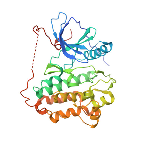
4TKS

Native-SAD phasing for human EGFR kinase domain.


Experimental Data Snapshot

  • Method: X-RAY DIFFRACTION
  • Resolution: 3.20 Å
  • R-Value Free: 0.209 
  • R-Value Work: 0.188 
  • R-Value Observed: 0.189 

wwPDB Validation   3D Report Full Report


This is version 1.2 of the entry. See complete history


Literature

Multi-crystal native SAD analysis at 6 keV.

Liu, Q.Guo, Y.Chang, Y.Cai, Z.Assur, Z.Mancia, F.Greene, M.I.Hendrickson, W.A.

(2014) Acta Crystallogr D Biol Crystallogr 70: 2544-2557

  • DOI: https://doi.org/10.1107/S1399004714013376
  • Primary Citation of Related Structures:  
    4TKQ, 4TKR, 4TKS

  • PubMed Abstract: 

    Anomalous diffraction signals from typical native macromolecules are very weak, frustrating their use in de novo structure determination. Here, native SAD procedures are described to enhance signal to noise in anomalous diffraction by using multiple crystals in combination with synchrotron X-rays at 6 keV. Increased anomalous signals were obtained at 6 keV compared with 7 keV X-ray energy, which was used for previous native SAD analyses. A feasibility test of multi-crystal-based native SAD phasing was performed at 3.2 Å resolution for a known tyrosine protein kinase domain, and real-life applications were made to two novel membrane proteins at about 3.0 Å resolution. The three applications collectively serve to validate the robust feasibility of native SAD phasing at lower energy.


  • Organizational Affiliation

    NYCOMPS, New York Structural Biology Center, New York, NY 10032, USA.


Macromolecules
Find similar proteins by:  (by identity cutoff)  |  3D Structure
Entity ID: 1
MoleculeChains Sequence LengthOrganismDetailsImage
Epidermal growth factor receptor332Homo sapiensMutation(s): 0 
Gene Names: EGFRERBBERBB1HER1
EC: 2.7.10.1
UniProt & NIH Common Fund Data Resources
Find proteins for P00533 (Homo sapiens)
Explore P00533 
Go to UniProtKB:  P00533
PHAROS:  P00533
GTEx:  ENSG00000146648 
Entity Groups  
Sequence Clusters30% Identity50% Identity70% Identity90% Identity95% Identity100% Identity
UniProt GroupP00533
Sequence Annotations
Expand
  • Reference Sequence
Experimental Data & Validation

Experimental Data

  • Method: X-RAY DIFFRACTION
  • Resolution: 3.20 Å
  • R-Value Free: 0.209 
  • R-Value Work: 0.188 
  • R-Value Observed: 0.189 
  • Space Group: I 2 3
Unit Cell:
Length ( Å )Angle ( ˚ )
a = 146.161α = 90
b = 146.161β = 90
c = 146.161γ = 90
Software Package:
Software NamePurpose
PHENIXrefinement

Structure Validation

View Full Validation Report


Entry History 

Deposition Data

Revision History  (Full details and data files)

  • Version 1.0: 2014-10-08
    Type: Initial release
  • Version 1.1: 2014-10-22
    Changes: Database references
  • Version 1.2: 2023-12-27
    Changes: Data collection, Database references, Derived calculations, Other, Refinement description, Source and taxonomy, Structure summary