4SBV | pdb_00004sbv

The REFINEMENT OF SOUTHERN BEAN MOSAIC VIRUS IN RECIPROCAL SPACE


Experimental Data Snapshot

  • Method: X-RAY DIFFRACTION
  • Resolution: 2.80 Å
  • R-Value Work: 
    0.254 (Depositor), 0.253 (DCC) 

wwPDB Validation 3D Report Full Report

Validation slider image for 4SBV

This is version 2.2 of the entry. See complete history

Literature

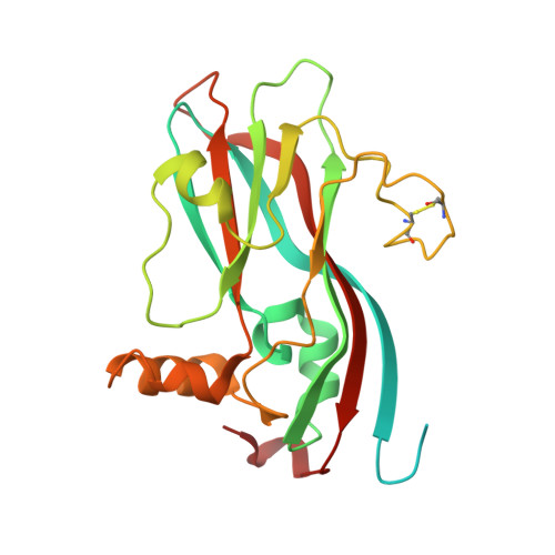
Refined structure of southern bean mosaic virus at 2.9 A resolution.

Silva, A.M.Rossmann, M.G.

(1987) J Mol Biology 197: 69-87

  • DOI: https://doi.org/10.1016/0022-2836(87)90610-3
  • Primary Citation Related Structures: 
    4SBV

  • PubMed Abstract: 

    The T = 3 capsid of southern bean mosaic virus is analyzed in detail. The beta-sheets of the beta-barrel folding motif that form the subunits show a high degree of twist, generated by several beta-bulges. Only 34 water molecules were identified in association with the three quasi-equivalent subunits, most of them on the external viral surface. Subunit contacts related by quasi-3-fold axes are similar, are dominated by polar interactions and have almost identical calcium binding sites. There is no metal ion on the quasi-3-fold axis, as previously reported. Subunits related by quasi-2-fold and icosahedral 2-fold axes have different contacts but nevertheless display almost identical interactions between the antiparallel helices alpha A. A dipole-dipole type interaction between these helices may produce an energetically stable hinge that allows two types of dimers in a T = 3 assembly. The temperature factor distribution, the hydrogen-bonding pattern, and the contacts across the icosahedral 2-fold axes suggest that one of the dimer types is present in the intact virion and probably also in solution; the other is produced only during capsid assembly. Interactions along the 5-fold axes are mainly polar and possibly form an ion channel. The beta-sheet structures of the three subunits can be superimposed with considerable precision. Significant relative distortions between quasi-equivalent subunits occur mainly in helices and loops. The two dimeric forms and the subunit distortions are the consequence of the non-equivalent subunit environments in the capsid.


  • Organizational Affiliation
    • Departamento de Fisica, Facultad de Ciencias Exactas, Universidad Nacional de La Plata, Argentina.

Macromolecule Content 

  • Total Structure Weight: 84.91 kDa 
  • Atom Count: 4,723 
  • Modeled Residue Count: 620 
  • Deposited Residue Count: 780 
  • Unique protein chains: 1

Macromolecules

Find similar proteins by:|  3D Structure
Entity ID: 1
MoleculeChains  Sequence LengthOrganismDetailsImage
SOUTHERN BEAN MOSAIC VIRUS COAT PROTEIN
A, B, C
260Southern bean mosaic virusMutation(s): 0 
UniProt
Find proteins for P03607 (Southern cowpea mosaic virus)
Explore P03607 
Go to UniProtKB:  P03607
Entity Groups
Sequence Clusters30% Identity50% Identity70% Identity90% Identity95% Identity100% Identity
UniProt GroupP03607
Sequence Annotations
Expand
Reference Sequence

Experimental Data & Validation

Experimental Data

  • Method: X-RAY DIFFRACTION
  • Resolution: 2.80 Å
  • R-Value Work:  0.254 (Depositor), 0.253 (DCC) 
Space Group: H 3 2
Unit Cell:
Length ( Å )Angle ( ˚ )
a = 334.3α = 90
b = 334.3β = 90
c = 757.5γ = 120

Structure Validation

View Full Validation Report


Entry History 

Deposition Data

  • Released Date: 1985-07-17 
  • Deposition Author(s): Rossmann, M.G.
  • This entry supersedes: 3SBV

Revision History  (Full details and data files)

  • Version 1.0: 1985-07-17
    Type: Initial release
  • Version 1.1: 2008-03-03
    Changes: Version format compliance
  • Version 1.2: 2011-07-13
    Changes: Version format compliance
  • Version 1.3: 2012-03-21
    Changes: Advisory
  • Version 2.0: 2023-04-19
    Type: Remediation
    Changes: Advisory, Atomic model, Data collection, Database references, Derived calculations, Other, Refinement description
  • Version 2.1: 2023-06-28
    Changes: Database references
  • Version 2.2: 2024-10-23
    Changes: Data collection, Structure summary