Novel mode of inhibition by D-tagatose 6-phosphate through a Heyns rearrangement in the active site of transaldolase B variants.
Stellmacher, L., Sandalova, T., Schneider, S., Schneider, G., Sprenger, G.A., Samland, A.K.(2016) Acta Crystallogr D Struct Biol 72: 467-476
- PubMed: 27050126 Search on PubMed
- DOI: https://doi.org/10.1107/S2059798316001170
- Primary Citation Related Structures:
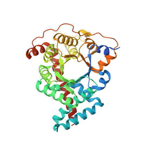
4S2B, 4S2C - PubMed Abstract:
Transaldolase B (TalB) and D-fructose-6-phosphate aldolase A (FSAA) from Escherichia coli are C-C bond-forming enzymes. Using kinetic inhibition studies and mass spectrometry, it is shown that enzyme variants of FSAA and TalB that exhibit D-fructose-6-phosphate aldolase activity are inhibited covalently and irreversibly by D-tagatose 6-phosphate (D-T6P), whereas no inhibition was observed for wild-type transaldolase B from E. coli. The crystal structure of the variant TalB(F178Y) with bound sugar phosphate was solved to a resolution of 1.46 Å and revealed a novel mode of covalent inhibition. The sugar is bound covalently via its C2 atom to the ℇ-NH2 group of the active-site residue Lys132. It is neither bound in the open-chain form nor as the closed-ring form of D-T6P, but has been converted to β-D-galactofuranose 6-phosphate (D-G6P), a five-membered ring structure. The furanose ring of the covalent adduct is formed via a Heyns rearrangement and subsequent hemiacetal formation. This reaction is facilitated by Tyr178, which is proposed to act as acid-base catalyst. The crystal structure of the inhibitor complex is compared with the structure of the Schiff-base intermediate of TalB(E96Q) formed with the substrate D-fructose 6-phosphate determined to a resolution of 2.20 Å. This comparison highlights the differences in stereochemistry at the C4 atom of the ligand as an essential determinant for the formation of the inhibitor adduct in the active site of the enzyme.
- Institut für Mikrobiologie, Universität Stuttgart, Allmandring 31, 70550 Stuttgart, Germany.
Organizational Affiliation: