4S0F | pdb_00004s0f

Crystal structure of the peptidase-containing ABC transporter PCAT1 E648Q mutant complexed with ATPgS in an occluded conformation


Experimental Data Snapshot

  • Method: X-RAY DIFFRACTION
  • Resolution: 5.51 Å
  • R-Value Free: 
    0.314 (Depositor), 0.333 (DCC) 
  • R-Value Work: 
    0.300 (Depositor), 0.332 (DCC) 
  • R-Value Observed: 
    0.301 (Depositor) 

Starting Models: experimental
View more details

wwPDB Validation 3D Report Full Report

Validation slider image for 4S0F

Ligand Structure Quality Assessment 


This is version 1.2 of the entry. See complete history

Literature

Crystal structures of a polypeptide processing and secretion transporter.

Lin, D.Y.Huang, S.Chen, J.

(2015) Nature 523: 425-430

  • DOI: https://doi.org/10.1038/nature14623
  • Primary Citation Related Structures: 
    4RY2, 4S0F

  • PubMed Abstract: 

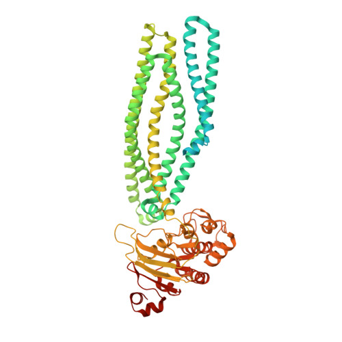
    Bacteria secrete peptides and proteins to communicate, to poison competitors, and to manipulate host cells. Among the various protein-translocation machineries, the peptidase-containing ATP-binding cassette transporters (PCATs) are appealingly simple. Each PCAT contains two peptidase domains that cleave the secretion signal from the substrate, two transmembrane domains that form a translocation pathway, and two nucleotide-binding domains that hydrolyse ATP. In Gram-positive bacteria, PCATs function both as maturation proteases and exporters for quorum-sensing or antimicrobial polypeptides. In Gram-negative bacteria, PCATs interact with two other membrane proteins to form the type 1 secretion system. Here we present crystal structures of PCAT1 from Clostridium thermocellum in two different conformations. These structures, accompanied by biochemical data, show that the translocation pathway is a large α-helical barrel sufficient to accommodate small folded proteins. ATP binding alternates access to the transmembrane pathway and also regulates the protease activity, thereby coupling substrate processing to translocation.


  • Organizational Affiliation
    • 1] Laboratory of Membrane Biology and Biophysics, The Rockefeller University, 1230 York Avenue, New York, New York 10065, USA [2] Howard Hughes Medical Institute, 1230 York Avenue, New York, New York 10065, USA.

Macromolecule Content 

  • Total Structure Weight: 163.41 kDa 
  • Atom Count: 5,636 
  • Modeled Residue Count: 1,130 
  • Deposited Residue Count: 1,460 
  • Unique protein chains: 1

Macromolecules

Find similar proteins by:|  3D Structure
Entity ID: 1
MoleculeChains  Sequence LengthOrganismDetailsImage
ABC-type bacteriocin transporter
A, B
730Acetivibrio thermocellus ATCC 27405Mutation(s): 1 
Gene Names: ABN51770.1Cthe_0534
EC: 3.4.22
Membrane Entity: Yes 
UniProt
Find proteins for A3DCU1 (Acetivibrio thermocellus (strain ATCC 27405 / DSM 1237 / JCM 9322 / NBRC 103400 / NCIMB 10682 / NRRL B-4536 / VPI 7372))
Explore A3DCU1 
Go to UniProtKB:  A3DCU1
Entity Groups
Sequence Clusters30% Identity50% Identity70% Identity90% Identity95% Identity100% Identity
UniProt GroupA3DCU1
Sequence Annotations
Expand
Reference Sequence

Small Molecules

Experimental Data & Validation

Experimental Data

  • Method: X-RAY DIFFRACTION
  • Resolution: 5.51 Å
  • R-Value Free:  0.314 (Depositor), 0.333 (DCC) 
  • R-Value Work:  0.300 (Depositor), 0.332 (DCC) 
  • R-Value Observed: 0.301 (Depositor) 
Space Group: P 42 21 2
Unit Cell:
Length ( Å )Angle ( ˚ )
a = 230α = 90
b = 230β = 90
c = 89.4γ = 90
Software Package:
Software NamePurpose
JBluIce-EPICSdata collection
PHASERphasing
PHENIXrefinement
HKL-2000data reduction
HKL-2000data scaling

Structure Validation

View Full Validation Report


Ligand Structure Quality Assessment 


Entry History 

Deposition Data

Revision History  (Full details and data files)

  • Version 1.0: 2015-07-22
    Type: Initial release
  • Version 1.1: 2015-08-05
    Changes: Database references
  • Version 1.2: 2023-09-20
    Changes: Data collection, Database references, Derived calculations, Refinement description