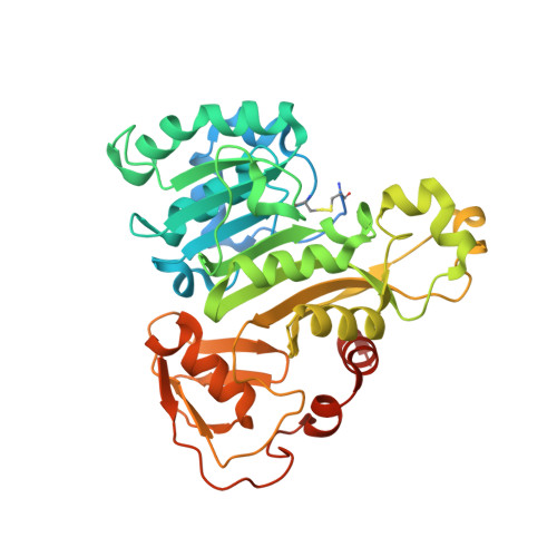
4RSR

ArsM arsenic(III) S-adenosylmethionine methyltransferase with trivalent phenyl arsencial derivative-Roxarsone


Experimental Data Snapshot

  • Method: X-RAY DIFFRACTION
  • Resolution: 2.25 Å
  • R-Value Free: 0.224 
  • R-Value Work: 0.173 
  • R-Value Observed: 0.176 

Starting Model: experimental
View more details

wwPDB Validation   3D Report Full Report


Ligand Structure Quality Assessment 


This is version 1.2 of the entry. See complete history


Literature

A disulfide-bond cascade mechanism for arsenic(III) S-adenosylmethionine methyltransferase.

Marapakala, K.Packianathan, C.Ajees, A.A.Dheeman, D.S.Sankaran, B.Kandavelu, P.Rosen, B.P.

(2015) Acta Crystallogr D Biol Crystallogr 71: 505-515

  • DOI: https://doi.org/10.1107/S1399004714027552
  • Primary Citation of Related Structures:  
    4KW7, 4RSR

  • PubMed Abstract: 

    Methylation of the toxic metalloid arsenic is widespread in nature. Members of every kingdom have arsenic(III) S-adenosylmethionine (SAM) methyltransferase enzymes, which are termed ArsM in microbes and AS3MT in animals, including humans. Trivalent arsenic(III) is methylated up to three times to form methylarsenite [MAs(III)], dimethylarsenite [DMAs(III)] and the volatile trimethylarsine [TMAs(III)]. In microbes, arsenic methylation is a detoxification process. In humans, MAs(III) and DMAs(III) are more toxic and carcinogenic than either inorganic arsenate or arsenite. Here, new crystal structures are reported of ArsM from the thermophilic eukaryotic alga Cyanidioschyzon sp. 5508 (CmArsM) with the bound aromatic arsenicals phenylarsenite [PhAs(III)] at 1.80 Å resolution and reduced roxarsone [Rox(III)] at 2.25 Å resolution. These organoarsenicals are bound to two of four conserved cysteine residues: Cys174 and Cys224. The electron density extends the structure to include a newly identified conserved cysteine residue, Cys44, which is disulfide-bonded to the fourth conserved cysteine residue, Cys72. A second disulfide bond between Cys72 and Cys174 had been observed previously in a structure with bound SAM. The loop containing Cys44 and Cys72 shifts by nearly 6.5 Å in the arsenic(III)-bound structures compared with the SAM-bound structure, which suggests that this movement leads to formation of the Cys72-Cys174 disulfide bond. A model is proposed for the catalytic mechanism of arsenic(III) SAM methyltransferases in which a disulfide-bond cascade maintains the products in the trivalent state.


  • Organizational Affiliation

    Department of Chemistry, Osmania University College for Women, Osmania University, Hyderabad 500 095, India.


Macromolecules
Find similar proteins by:  (by identity cutoff)  |  3D Structure
Entity ID: 1
MoleculeChains Sequence LengthOrganismDetailsImage
Arsenic methyltransferase383Cyanidioschyzon sp. 5508Mutation(s): 0 
Gene Names: arsM
UniProt
Find proteins for C0JV69 (Cyanidioschyzon sp. 5508)
Explore C0JV69 
Go to UniProtKB:  C0JV69
Entity Groups  
Sequence Clusters30% Identity50% Identity70% Identity90% Identity95% Identity100% Identity
UniProt GroupC0JV69
Sequence Annotations
Expand
  • Reference Sequence
Small Molecules
Ligands 5 Unique
IDChains Name / Formula / InChI Key2D Diagram3D Interactions
RXO
Query on RXO

Download Ideal Coordinates CCD File 
B [auth A]4-arsanyl-2-nitrophenol
C6 H6 As N O3
POYWCBSQXBGKKH-UHFFFAOYSA-N
DTT
Query on DTT

Download Ideal Coordinates CCD File 
G [auth A]2,3-DIHYDROXY-1,4-DITHIOBUTANE
C4 H10 O2 S2
VHJLVAABSRFDPM-IMJSIDKUSA-N
CA
Query on CA

Download Ideal Coordinates CCD File 
D [auth A],
E [auth A]
CALCIUM ION
Ca
BHPQYMZQTOCNFJ-UHFFFAOYSA-N
MG
Query on MG

Download Ideal Coordinates CCD File 
C [auth A]MAGNESIUM ION
Mg
JLVVSXFLKOJNIY-UHFFFAOYSA-N
NA
Query on NA

Download Ideal Coordinates CCD File 
F [auth A]SODIUM ION
Na
FKNQFGJONOIPTF-UHFFFAOYSA-N
Experimental Data & Validation

Experimental Data

  • Method: X-RAY DIFFRACTION
  • Resolution: 2.25 Å
  • R-Value Free: 0.224 
  • R-Value Work: 0.173 
  • R-Value Observed: 0.176 
  • Space Group: C 1 2 1
Unit Cell:
Length ( Å )Angle ( ˚ )
a = 85.156α = 90
b = 46.406β = 114.07
c = 100.255γ = 90
Software Package:
Software NamePurpose
SERGUIdata collection
PHASERphasing
REFMACrefinement
HKL-2000data reduction
HKL-2000data scaling

Structure Validation

View Full Validation Report


Ligand Structure Quality Assessment 


Entry History 

Deposition Data

Revision History  (Full details and data files)

  • Version 1.0: 2014-12-10
    Type: Initial release
  • Version 1.1: 2015-03-18
    Changes: Database references
  • Version 1.2: 2023-09-20
    Changes: Data collection, Database references, Derived calculations, Refinement description