4RS7 | pdb_00004rs7

Structure of pNOB8 ParB-C


Experimental Data Snapshot

  • Method: X-RAY DIFFRACTION
  • Resolution: 1.90 Å
  • R-Value Free: 
    0.224 (Depositor), 0.194 (DCC) 
  • R-Value Work: 
    0.193 (Depositor), 0.191 (DCC) 
  • R-Value Observed: 
    0.195 (Depositor) 

wwPDB Validation   3D Report Full Report


This is version 1.1 of the entry. See complete history


Literature

Structures of archaeal DNA segregation machinery reveal bacterial and eukaryotic linkages.

Schumacher, M.A.Tonthat, N.K.Lee, J.Rodriguez-Castaneda, F.A.Chinnam, N.B.Kalliomaa-Sanford, A.K.Ng, I.W.Barge, M.T.Shaw, P.L.Barilla, D.

(2015) Science 349: 1120-1124

  • DOI: https://doi.org/10.1126/science.aaa9046
  • Primary Citation of Related Structures:  
    4RS7, 4RS8, 5K5A, 5K5D, 5K5O, 5K5Q, 5K5R, 5K5Z

  • PubMed Abstract: 

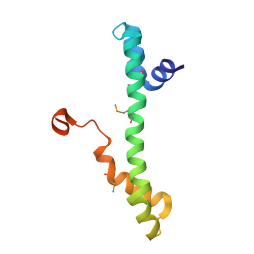
    Although recent studies have provided a wealth of information about archaeal biology, nothing is known about the molecular basis of DNA segregation in these organisms. Here, we unveil the machinery and assembly mechanism of the archaeal Sulfolobus pNOB8 partition system. This system uses three proteins: ParA; an atypical ParB adaptor; and a centromere-binding component, AspA. AspA utilizes a spreading mechanism to create a DNA superhelix onto which ParB assembles. This supercomplex links to the ParA motor, which contains a bacteria-like Walker motif. The C domain of ParB harbors structural similarity to CenpA, which dictates eukaryotic segregation. Thus, this archaeal system combines bacteria-like and eukarya-like components, which suggests the possible conservation of DNA segregation principles across the three domains of life.


  • Organizational Affiliation
    • Department of Biochemistry, Duke University School of Medicine, 243 Nanaline H. Duke, Box 3711, Durham, NC 27710, USA. maria.schumacher@duke.edu daniela.barilla@york.ac.uk.

Macromolecules
Find similar proteins by:  (by identity cutoff)  |  3D Structure
Entity ID: 1
MoleculeChains Sequence LengthOrganismDetailsImage
ParB-CA [auth R],
B [auth D],
C [auth A],
D [auth B]
96Sulfolobus sp. NOB8H2Mutation(s): 0 
Entity Groups  
Sequence Clusters30% Identity50% Identity70% Identity90% Identity95% Identity100% Identity
Sequence Annotations
Expand
  • Reference Sequence
Small Molecules
Modified Residues  1 Unique
IDChains TypeFormula2D DiagramParent
MSE
Query on MSE
A [auth R],
B [auth D],
C [auth A],
D [auth B]
L-PEPTIDE LINKINGC5 H11 N O2 SeMET
Experimental Data & Validation

Experimental Data

  • Method: X-RAY DIFFRACTION
  • Resolution: 1.90 Å
  • R-Value Free:  0.224 (Depositor), 0.194 (DCC) 
  • R-Value Work:  0.193 (Depositor), 0.191 (DCC) 
  • R-Value Observed: 0.195 (Depositor) 
Space Group: P 1 21 1
Unit Cell:
Length ( Å )Angle ( ˚ )
a = 50.21α = 90
b = 56.06β = 94.4
c = 72.3γ = 90
Software Package:
Software NamePurpose
ADSCdata collection
SOLVEphasing
PHENIXrefinement
MOSFLMdata reduction
SCALAdata scaling

Structure Validation

View Full Validation Report


Entry History 

Deposition Data

Revision History  (Full details and data files)

  • Version 1.0: 2015-09-16
    Type: Initial release
  • Version 1.1: 2024-10-16
    Changes: Data collection, Database references, Derived calculations, Structure summary