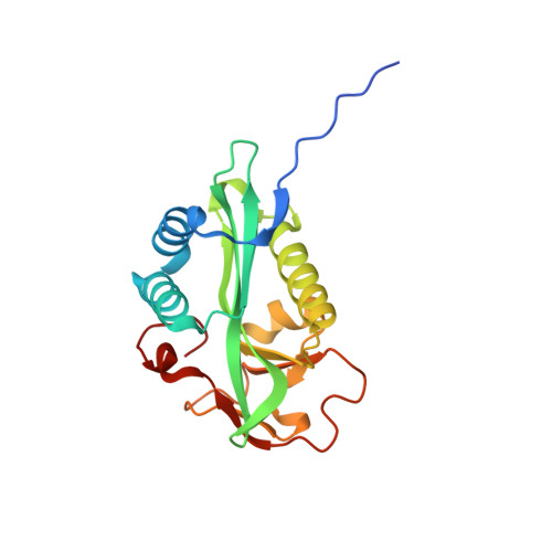
1.55 Angstrom Crystal Structure of GNAT Family N-acetyltransferase (YhbS) from Escherichia coli in Complex with CoA.
Minasov, G., Wawrzak, Z., Kuhn, M., Shuvalova, L., Dubrovska, I., Flores, K., Anderson, W.F., Center for Structural Genomics of Infectious Diseases (CSGID)To be published.