4RRZ

Crystal Structure of Apo Murine H90W Cyclooxygenase-2 Complexed with Lumiracoxib


Experimental Data Snapshot

  • Method: X-RAY DIFFRACTION
  • Resolution: 2.57 Å
  • R-Value Free: 0.216 
  • R-Value Work: 0.184 
  • R-Value Observed: 0.185 

wwPDB Validation   3D Report Full Report


Ligand Structure Quality Assessment 


This is version 2.1 of the entry. See complete history


Literature

Action at a Distance: MUTATIONS OF PERIPHERAL RESIDUES TRANSFORM RAPID REVERSIBLE INHIBITORS TO SLOW, TIGHT BINDERS OF CYCLOOXYGENASE-2.

Blobaum, A.L.Xu, S.Rowlinson, S.W.Duggan, K.C.Banerjee, S.Kudalkar, S.N.Birmingham, W.R.Ghebreselasie, K.Marnett, L.J.

(2015) J Biol Chem 290: 12793-12803

  • DOI: https://doi.org/10.1074/jbc.M114.635987
  • Primary Citation of Related Structures:  
    4RRW, 4RRX, 4RRY, 4RRZ, 4RS0

  • PubMed Abstract: 

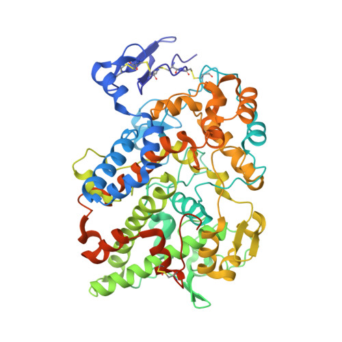
    Cyclooxygenase enzymes (COX-1 and COX-2) catalyze the conversion of arachidonic acid to prostaglandin G2. The inhibitory activity of rapid, reversible COX inhibitors (ibuprofen, naproxen, mefenamic acid, and lumiracoxib) demonstrated a significant increase in potency and time dependence of inhibition against double tryptophan murine COX-2 mutants at the 89/90 and 89/119 positions. In contrast, the slow, time-dependent COX inhibitors (diclofenac, indomethacin, and flurbiprofen) were unaffected by those mutations. Further mutagenesis studies suggested that mutation at position 89 was principally responsible for the changes in inhibitory potency of rapid, reversible inhibitors, whereas mutation at position 90 may exert some effect on the potency of COX-2-selective diarylheterocycle inhibitors; no effect was observed with mutation at position 119. Several crystal structures with or without NSAIDs indicated that placement of a bulky residue at position 89 caused a closure of a gap at the lobby, and alteration of histidine to tryptophan at position 90 changed the electrostatic profile of the side pocket of COX-2. Thus, these two residues, especially Val-89 at the lobby region, are crucial for the entrance and exit of some NSAIDs from the COX active site.


  • Organizational Affiliation

    From the A. B. Hancock, Jr. Memorial Laboratory for Cancer Research, Departments of Biochemistry, Chemistry, and Pharmacology, Vanderbilt Institute of Chemical Biology, Center in Molecular Toxicology, Vanderbilt-Ingram Cancer Center, Vanderbilt University School of Medicine, Nashville Tennessee 37232.


Macromolecules
Find similar proteins by:  (by identity cutoff)  |  3D Structure
Entity ID: 1
MoleculeChains Sequence LengthOrganismDetailsImage
Prostaglandin G/H synthase 2
A, B, C, D
587Mus musculusMutation(s): 1 
Gene Names: Ptgs2Cox-2Cox2Pghs-bTis10
EC: 1.14.99.1
UniProt
Find proteins for Q05769 (Mus musculus)
Explore Q05769 
Go to UniProtKB:  Q05769
Entity Groups  
Sequence Clusters30% Identity50% Identity70% Identity90% Identity95% Identity100% Identity
UniProt GroupQ05769
Sequence Annotations
Expand
  • Reference Sequence
Oligosaccharides

Help

Entity ID: 2
MoleculeChains Length2D Diagram Glycosylation3D Interactions
2-acetamido-2-deoxy-beta-D-glucopyranose-(1-4)-2-acetamido-2-deoxy-beta-D-glucopyranose
E, F, G, H
2N-Glycosylation
Glycosylation Resources
GlyTouCan:  G42666HT
GlyCosmos:  G42666HT
GlyGen:  G42666HT
Small Molecules
Ligands 2 Unique
IDChains Name / Formula / InChI Key2D Diagram3D Interactions
LUR
Query on LUR

Download Ideal Coordinates CCD File 
K [auth A],
N [auth B],
Q [auth C],
T [auth D]
{2-[(2-chloro-6-fluorophenyl)amino]-5-methylphenyl}acetic acid
C15 H13 Cl F N O2
KHPKQFYUPIUARC-UHFFFAOYSA-N
NAG
Query on NAG

Download Ideal Coordinates CCD File 
I [auth A]
J [auth A]
L [auth B]
M [auth B]
O [auth C]
I [auth A],
J [auth A],
L [auth B],
M [auth B],
O [auth C],
P [auth C],
R [auth D],
S [auth D]
2-acetamido-2-deoxy-beta-D-glucopyranose
C8 H15 N O6
OVRNDRQMDRJTHS-FMDGEEDCSA-N
Binding Affinity Annotations 
IDSourceBinding Affinity
LUR BindingDB:  4RRZ Ki: 320 (nM) from 1 assay(s)
IC50: min: 7, max: 1.00e+4 (nM) from 7 assay(s)
Experimental Data & Validation

Experimental Data

  • Method: X-RAY DIFFRACTION
  • Resolution: 2.57 Å
  • R-Value Free: 0.216 
  • R-Value Work: 0.184 
  • R-Value Observed: 0.185 
  • Space Group: C 1 2 1
Unit Cell:
Length ( Å )Angle ( ˚ )
a = 215.711α = 90
b = 121.047β = 123.2
c = 135.111γ = 90
Software Package:
Software NamePurpose
HKL-2000data collection
PHASESphasing
PHENIXrefinement
XDSdata reduction
XDSdata scaling

Structure Validation

View Full Validation Report


Ligand Structure Quality Assessment 


Entry History 

Deposition Data

Revision History  (Full details and data files)

  • Version 1.0: 2015-04-08
    Type: Initial release
  • Version 1.1: 2015-05-06
    Changes: Database references
  • Version 1.2: 2015-06-03
    Changes: Database references
  • Version 2.0: 2020-07-29
    Type: Remediation
    Reason: Carbohydrate remediation
    Changes: Atomic model, Data collection, Database references, Derived calculations, Structure summary
  • Version 2.1: 2023-09-20
    Changes: Data collection, Database references, Refinement description, Structure summary