4RMD | pdb_00004rmd

Crystal structure of human Retinoid X receptor alpha ligand binding domain complex with 9cUAB110 and coactivator peptide GRIP-1


Experimental Data Snapshot

  • Method: X-RAY DIFFRACTION
  • Resolution: 1.90 Å
  • R-Value Free: 
    0.239 (Depositor), 0.213 (DCC) 
  • R-Value Work: 
    0.209 (Depositor), 0.208 (DCC) 
  • R-Value Observed: 
    0.209 (Depositor) 

Starting Model: experimental
View more details

wwPDB Validation 3D Report Full Report

Validation slider image for 4RMD

Ligand Structure Quality Assessment 


This is version 1.3 of the entry. See complete history

Literature

Conformationally Defined Rexinoids and Their Efficacy in the Prevention of Mammary Cancers.

Atigadda, V.R.Xia, G.Deshpande, A.Wu, L.Kedishvili, N.Smith, C.D.Krontiras, H.Bland, K.I.Grubbs, C.J.Brouillette, W.J.Muccio, D.D.

(2015) J Med Chem 58: 7763-7774

  • DOI: https://doi.org/10.1021/acs.jmedchem.5b00829
  • Primary Citation Related Structures: 
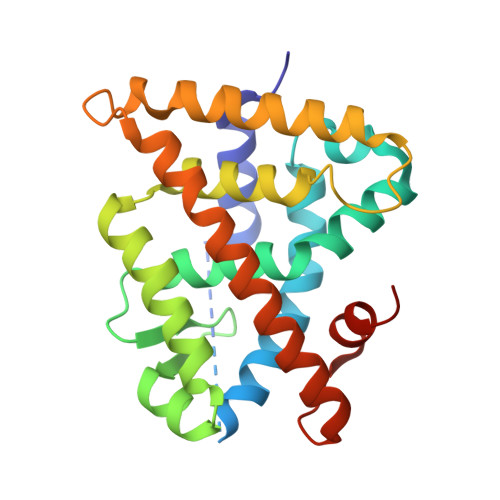
    4RFW, 4RMC, 4RMD, 4RME

  • PubMed Abstract: 

    (2E,4E,6Z,8Z)-8-(3',4'-Dihydro-1'(2H)-naphthalen-1'-ylidene)-3,7-dimethyl-2,3,6-octatrienoinic acid (UAB30) is currently undergoing clinical evaluation as a novel cancer prevention agent. In efforts to develop even more highly potent rexinoids that prevent breast cancer without toxicity, we further explore here the structure-activity relationship of two separate classes of rexinoids. UAB30 belongs to the class II rexinoids and possesses a 9Z-tetraenoic acid chain bonded to a tetralone ring, whereas the class I rexinoids contain the same 9Z-tetraenoic acid chain bonded to a disubstituted cyclohexenyl ring. Among the 12 class I and class II rexinoids evaluated, the class I rexinoid 11 is most effective in preventing breast cancers in an in vivo rat model alone or in combination with tamoxifen. Rexinoid 11 also reduces the size of established tumors and exhibits a therapeutic effect. However, 11 induces hypertriglyceridemia at its effective dose. On the other hand rexinoid 10 does not increase triglyceride levels while being effective in the in vivo chemoprevention assay. X-ray studies of four rexinoids bound to the ligand binding domain of the retinoid X receptor reveal key structural aspects that enhance potency as well as those that enhance the synthesis of lipids.


  • Organizational Affiliation
    • Departments of †Chemistry, ‡Biochemistry and Molecular Genetics, §Vision Sciences, and ∥Surgery, University of Alabama at Birmingham , Birmingham, Alabama 35294, United States.

Macromolecule Content 

  • Total Structure Weight: 28.33 kDa 
  • Atom Count: 1,937 
  • Modeled Residue Count: 225 
  • Deposited Residue Count: 248 
  • Unique protein chains: 2

Macromolecules

Find similar proteins by:|  3D Structure
Entity ID: 1
MoleculeChains  Sequence LengthOrganismDetailsImage
Retinoic acid receptor RXR-alpha235Homo sapiensMutation(s): 0 
Gene Names: NR2B1RXRARXRA NR2B1
UniProt & NIH Common Fund Data Resources
Find proteins for P19793 (Homo sapiens)
Explore P19793 
Go to UniProtKB:  P19793
PHAROS:  P19793
GTEx:  ENSG00000186350 
Entity Groups
Sequence Clusters30% Identity50% Identity70% Identity90% Identity95% Identity100% Identity
UniProt GroupP19793
Sequence Annotations
Expand
Reference Sequence
Find similar proteins by:  Sequence   |   3D Structure  
Entity ID: 2
MoleculeChains  Sequence LengthOrganismDetailsImage
Nuclear receptor coactivator 213Homo sapiensMutation(s): 0 
UniProt & NIH Common Fund Data Resources
Find proteins for Q15596 (Homo sapiens)
Explore Q15596 
Go to UniProtKB:  Q15596
PHAROS:  Q15596
GTEx:  ENSG00000140396 
Entity Groups
Sequence Clusters30% Identity50% Identity70% Identity90% Identity95% Identity100% Identity
UniProt GroupQ15596
Sequence Annotations
Expand
Reference Sequence

Small Molecules

Ligands 1 Unique
IDChains Name / Formula / InChI Key2D Diagram3D Interactions
3SW

Query on 3SW


Download:Ideal Coordinates CCD File
C [auth A](2E,4E,6Z,8E)-8-[3-cyclopropyl-2-(3-methylbutyl)cyclohex-2-en-1-ylidene]-3,7-dimethylocta-2,4,6-trienoic acid
C24 H34 O2
SCCVQQDOUFNKGM-RBEOCVBDSA-N
Binding Affinity Annotations 
IDSourceBinding Affinity
3SW BindingDB:  4RMD Kd: 35 (nM) from 1 assay(s)
EC50: 19 (nM) from 1 assay(s)

Experimental Data & Validation

Experimental Data

  • Method: X-RAY DIFFRACTION
  • Resolution: 1.90 Å
  • R-Value Free:  0.239 (Depositor), 0.213 (DCC) 
  • R-Value Work:  0.209 (Depositor), 0.208 (DCC) 
  • R-Value Observed: 0.209 (Depositor) 
Space Group: P 43 21 2
Unit Cell:
Length ( Å )Angle ( ˚ )
a = 65.971α = 90
b = 65.971β = 90
c = 112.347γ = 90
Software Package:
Software NamePurpose
HKL-2000data collection
CNSrefinement
DENZOdata reduction
HKL-2000data scaling
CNSphasing

Structure Validation

View Full Validation Report


Ligand Structure Quality Assessment 


Entry History 

Deposition Data

Revision History  (Full details and data files)

  • Version 1.0: 2015-09-23
    Type: Initial release
  • Version 1.1: 2015-10-21
    Changes: Database references
  • Version 1.2: 2022-08-17
    Changes: Database references, Derived calculations
  • Version 1.3: 2023-09-20
    Changes: Data collection, Refinement description